
Fig. S6
- ID
- ZDB-IMAGE-141124-37
- Genes
- Publication
- McGraw et al., 2014 - Kremen1 restricts Dkk activity during posterior lateral line development in zebrafish
- All Figures
- Figures for McGraw et al., 2014
|
Fig. S6
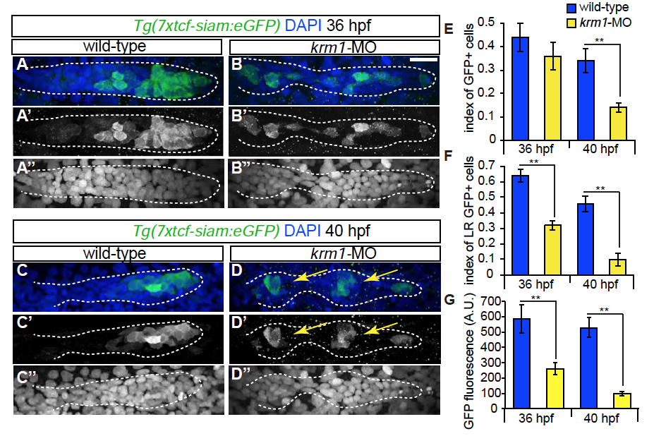
Wnt activity is decreased following Krm1 knockdown. (A-D′′′) Confocal projections of wild-type and kremen1 morphant embryos expressing the Wnt sensor Tg(7xtcf-siam:eGFP) (green); nuclei are labeled with DAPI (blue). (A-A′′, C-C′′) GFP is expressed in cells of the leading part of a wildtype pLLP at 36 and 40 hpf. (B-B′′,D-D′′) In kremen1-MO injected embryos, GFP-positive cells are located throughout the pLLP, most are incorporated into proto-NMs (yellow arrows) and notably reduced in the leading region. By 40 hpf, the index of GFP-positive cells is significantly reduced in kremen1 morphants (E). (F) The index of GFP in the leading zone of the pLLP (the caudal-most 30 cells of the pLLP) is significantly reduced in kremen1 morphants at 36 and 40 hpf as compared to wild-type controls (n=10-12 embryos/condition; **p<0.001 Student’s t-test). Scale bars=20μm. (G) The mean fluorescence intensity of GFP-positive cells, measured in arbitrary units (A.U.), was significantly reduced in morphant primordia at both 36 and 40 hpf (n=10-12 embryos/condition; **p<0.001 Student’s t-test).