
Fig. S5
- ID
- ZDB-IMAGE-141124-13
- Publication
- Schindler et al., 2014 - Hand2 elevates cardiomyocyte production during zebrafish heart development and regeneration
- All Figures
- Figures for Schindler et al., 2014
|
Fig. S5
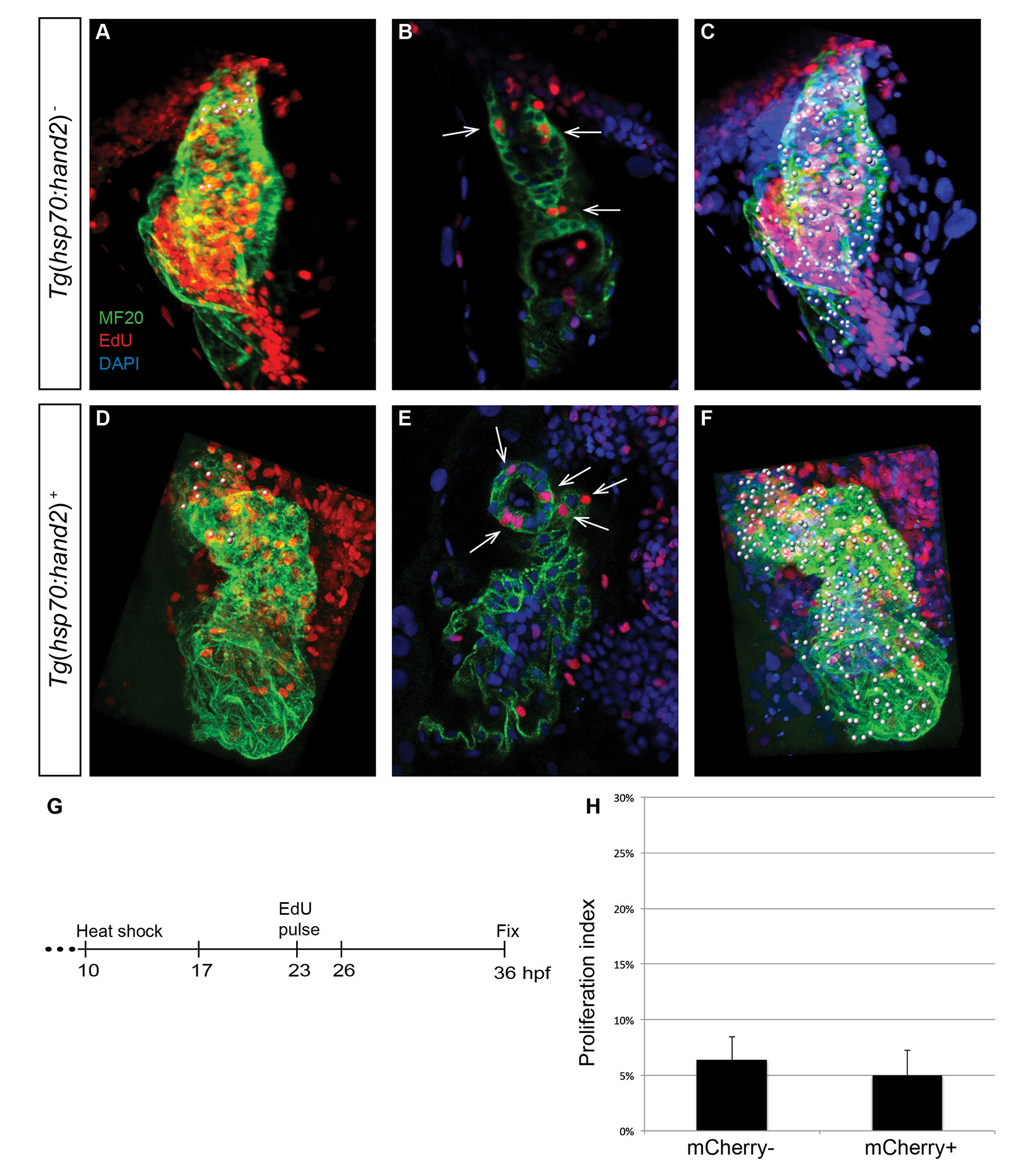
No evident influence of hand2 overexpression on cardiomyocyte proliferation after initial heart tube assembly. (A-F) EdU incorporation in hearts of (A-C) nontransgenic and (D-F) Tg(hsp70:hand2) embryos at 36 hpf, following heat shock at 10 hpf and EdU pulse at 23 hpf; (A,C,D,F) partial reconstructions of confocal z-stacks with ventricle up and (B,E) representative single slices. Dots, arrows, and color schemes are as described for Fig. 5A-F. Note that the nontransgenic heart shown (A) contains a number of EdU-positive blood cells that were trapped during fixation; EdU-positive blood cells are less commonly observed within the hearts of hand2-overexpressing embryos (D), due to their impaired circulation. (G) Timeline of experimental design. (H) Bar graph compares proliferation indexes in nontransgenic (mCherry-negative) and Tg(hsp70:hand2) (mCherry-positive) embryos, as in Fig. 5H. No change in proliferation index is seen in hand2-overexpressing embryos (n=8-11; p=0.196).