
Fig. 10
- ID
- ZDB-IMAGE-141107-4
- Publication
- Ichimura et al., 2013 - Podocalyxin regulates pronephric glomerular development in zebrafish
- All Figures
- Figures for Ichimura et al., 2013
|
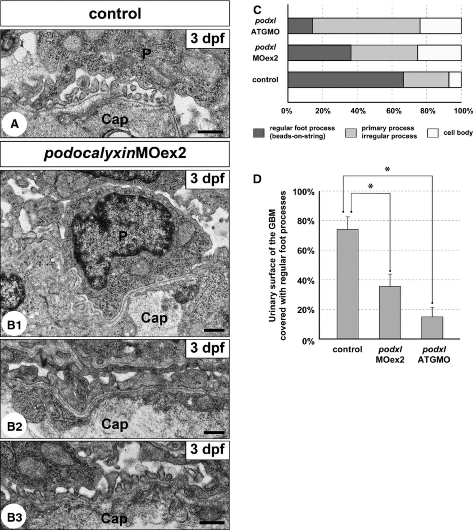
Fig. 10 Truncation of podocalyxin exon 2 induces poor development of podocyte foot processes. (A and B1-3) Transmission electron micrographs showing glomerular capillary wall. Unlike control larvae (A), podocyte cell bodies that directly adhere to the GBM (B1) and irregular-shaped processes that cover the GBM (B2) are frequently found in the podxlMOex2 morphants, although regular foot processes with a “beads-on-a-string” pattern are also formed in some regions (B3). Cap, glomerular capillary lumen; P, podocyte cell body. Bar scales: 500 nm. (C) The ratio of the three structures covering the urinary surface of GBM. (D) Significant reduction in regular foot processes with slit diaphragm in the podxlATGMO and podxlMOex2 morphants. In control larvae, regular foot processes with slit diaphragm cover 66.7 ± 7.8% of the urinary surface of the GBM (N = 3). The foot process-covering area is greatly reduced in the podxlMOex2 morphants (36.3 ± 5.0%, N = 3) and non-cystic-type podxlATGMO morphants (14.4 ± 7.5%, N = 3) with statistical significance. All of the larvae were examined on 3 dpf. *P < 0.01.