
Image
|
Figure Caption
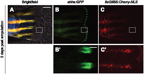
Fig. 4
Visualization of Hh signaling during adult fin regeneration.
Caudal fin of a Tg(-2.4shha:GFP-ABC;8xGliBS:mCherry-NLS) zebrafish three days after amputation. Brightfield (A) and fluorescence micrographs (B–C) are shown, with the dashed boxes corresponding to the magnified views below. Fin orientations: lateral views, anterior left. Scale bars: A–C, 2 mm; B2–C2, 200 μm.
Acknowledgments
This image is the copyrighted work of the attributed author or publisher, and
ZFIN has permission only to display this image to its users.
Additional permissions should be obtained from the applicable author or publisher of the image.
Full text @ PLoS One