
IMAGE
Fig. 5
- ID
- ZDB-IMAGE-140923-18
- Genes
- Antibodies
- Publication
- Lin et al., 2014 - Hypoxia-Inducible Factor 2 Alpha Is Essential for Hepatic Outgrowth and Functions via the Regulation of leg1 Transcription in the Zebrafish Embryo
- All Figures
- Figures for Lin et al., 2014
Image
|
Figure Caption
Fig. 5
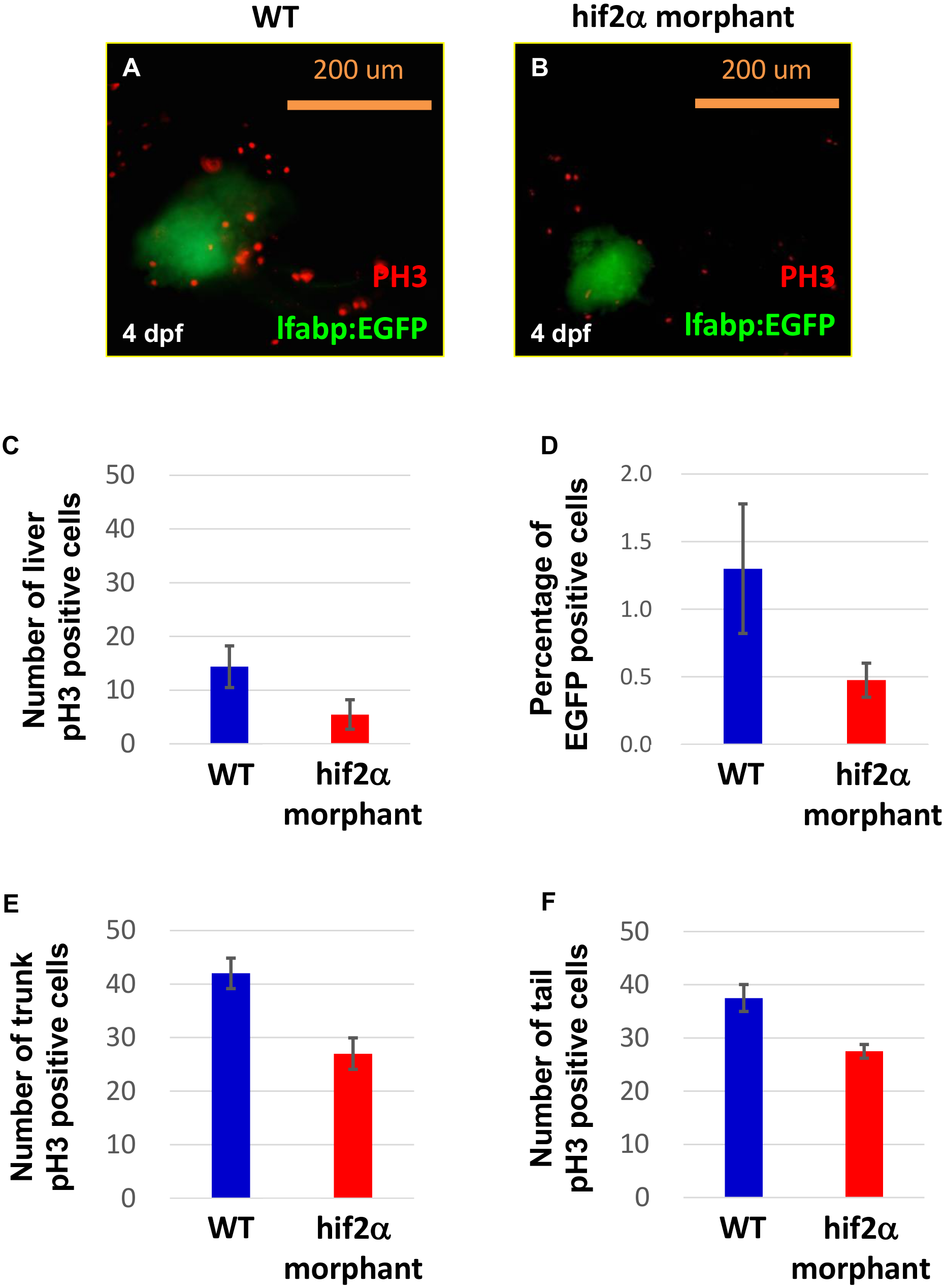
Knockdown of hif2-alpha damages liver cell proliferation.
WT (Tg(lfabp:EGFP)) embryos (A) and hif2-alpha ATG-MO-injected embryos (B) were examined for liver cell proliferation using an anti-pH3 antibody at 4 dpf. Cell proliferation in hif2-alpha ATG-MO-injected embryos was reduced compared with WT embryos. (C) Quantification of pH3-positive cells in the liver (n = 11, p<0.05). (D) The EGFP-positive cells were counted by FACS. Quantification of pH3-positive cells in the trunk (E) and tail (F) (n = 4, p<0.05).
Figure Data
Acknowledgments
This image is the copyrighted work of the attributed author or publisher, and
ZFIN has permission only to display this image to its users.
Additional permissions should be obtained from the applicable author or publisher of the image.
Full text @ PLoS One