
IMAGE
Fig. S1
- ID
- ZDB-IMAGE-140910-12
- Publication
- Xue et al., 2014 - Organizer-derived Bmp2 is required for the formation of a correct Bmp activity gradient during embryonic development
- All Figures
- Figures for Xue et al., 2014
Image
|
Figure Caption
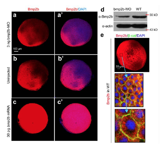
Fig. S1
Effectiveness of anti-Bmp2b antibody for detection of endogenous Bmp2b in zebrafish embryos. (a-c′) Immunofluorescence detection of endogenous Bmp2b.
Embryos were injected at the one-cell stage with 3 ng bmp2b-MO or 30 pg bmp2b mRNA and immunostained at the shield stage using anti-bmp2b antibody along with DAPI staining. All embryos were placed with animal pole to the top. Note that the signal was weakened in bmp2b morphant and greatly enhanced in bmp2b mRNA-injected embryo. (d) Bmp2b protein detection by western blotting. Embryos injected with 3 ng bmp2b-MO and uninjected embryos were harvested at the 75% epiboly stage and lysed for western blotting using anti-Bmp2b and anti-actin antibodies. Note that Bmp2b was reduced in bmp2b morphants. (e) Subcellular localization of Bmp2b proteins. A boxed area in the ventral region of the shield stage embryo (same one as shown in Fig. 1g) was enlarged in the middle picture and a single entire cell was further enlarged in the bottom picture.
Acknowledgments
This image is the copyrighted work of the attributed author or publisher, and
ZFIN has permission only to display this image to its users.
Additional permissions should be obtained from the applicable author or publisher of the image.
Full text @ Nat. Commun.