Image
Figure Caption
Fig. 4
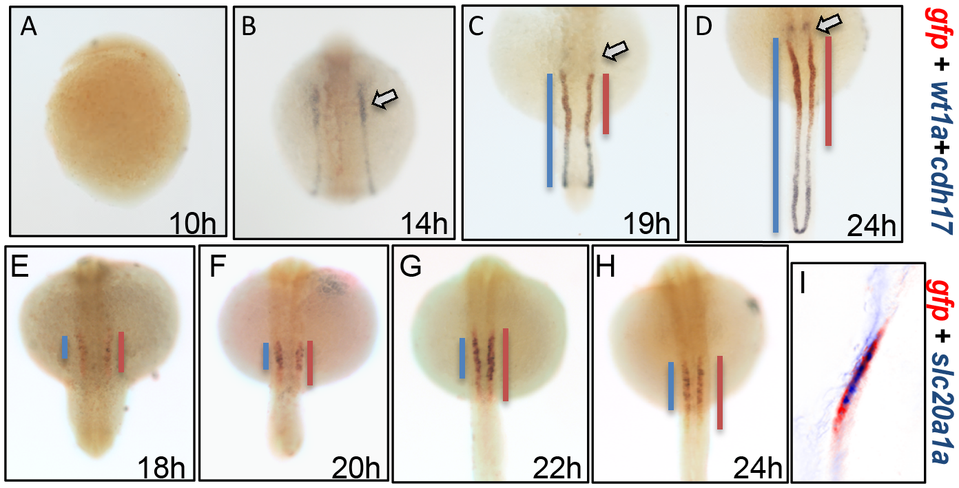
The gtshβ promoter-driven GFP is expressed in pronephric tubules.
(A–D) dWISH analyses of gfp (red), wt1a1 (purple) and cdh17 (purple) in Tg(gtshβ::GFP) embryos at the somitogenic stages from 10 hpf to 24 hpf. (E–H) dWISH analyses of gfp (red) and slc20a1a (purple) in Tg(gtshβ::GFP) embryos at the somitogenic stages from 18 hpf to 24 hpf. (I) Nuance multispectral imaging of gfp-slc20a1a-double-stained Tg(gtshβ::GFP) embryos at 24 hpf. The podocytes stained with wt1a1 are indicated by arrows. The blue or red lines are drawn according to the expression patterns of the kidney tubule markers.
Acknowledgments
This image is the copyrighted work of the attributed author or publisher, and
ZFIN has permission only to display this image to its users.
Additional permissions should be obtained from the applicable author or publisher of the image.
Full text @ PLoS One

