Image
Figure Caption
Fig. S5
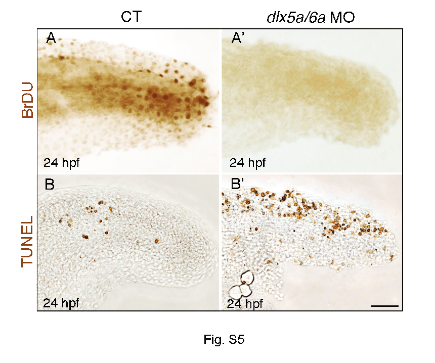
The median fin fold defects in dlx5a/6a morphants are associated with altered cell proliferation and apoptosis. Lateral view at the posterior axis of BrdU (A, A2) and TUNEL (B, B2) assays on control (A–B) and dlx5a/6a morphant (A2–B2) embryos at 24 hpf. The BrdU assay shows that controls present proliferating cells in the median fin fold whereas no BrdU-positive cells are observed in the morphants. In parallel, the morphants show a high increase of apoptotic cells in the MFF compared to controls (B–B2). Scale bar shown in B2 for all panels 20 μm.
Acknowledgments
This image is the copyrighted work of the attributed author or publisher, and
ZFIN has permission only to display this image to its users.
Additional permissions should be obtained from the applicable author or publisher of the image.
Full text @ PLoS One

