
Fig. S1
- ID
- ZDB-IMAGE-140902-7
- Publication
- Al-Hamed et al., 2014 - Functional modelling of a novel mutation in BBS5
- All Figures
- Figures for Al-Hamed et al., 2014
|
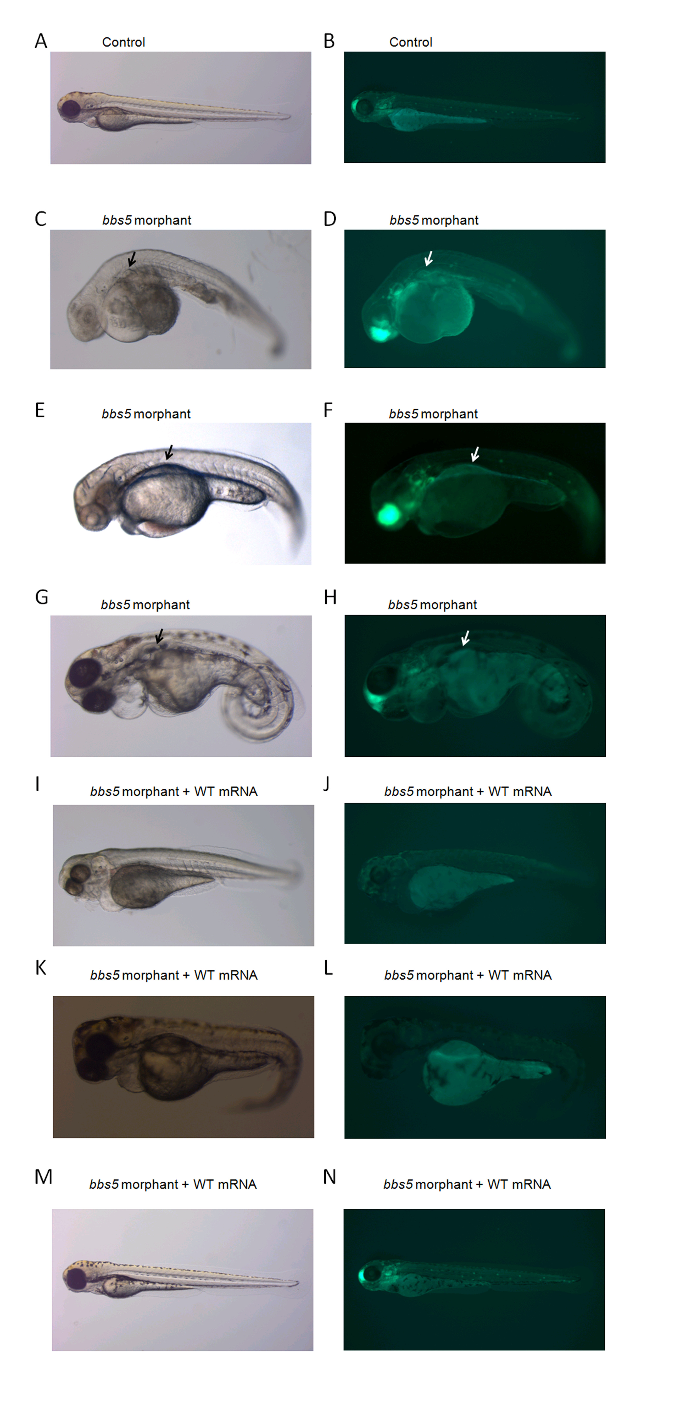
Fig. S1
Light and fluorescence microscopy of renal cysts in bbs5 morphants and rescue with WT bbs5 mRNA. Left panels show bright-field images of 72 hpf embryos and right panels show immunofluorescence images, using claudin-Lyn-GFP embryos which express GFP throughout the pronephros (as well as forebrain and ear). (A,B) Uninjected fish (Control). (C-H) Morphological defects are seen in bb5 morphant embryos. bbs5 morphant embryos show pronephros dilatation and cyst formation which is subtle on light microscopy (black arrows) but more easily identified under fluorescence microscopy (white arrows). (I-N) Morphant phenotypes of tail abnormalities, pronephric duct dilatation /cysts are (I-L) partially and (M,N) fully rescued by co-injection with WT bbs5 mRNA.