
Fig. 2
- ID
- ZDB-IMAGE-140902-30
- Publication
- Xue et al., 2014 - Organizer-derived Bmp2 is required for the formation of a correct Bmp activity gradient during embryonic development
- All Figures
- Figures for Xue et al., 2014
|
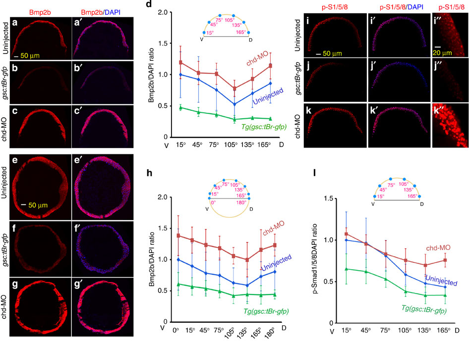
Fig. 2
(a–d) Changes in Bmp2b protein distribution on meridional sections of differently treated embryos. Meridional cryostat sections passing the organizer of WT (a,a2), Tg(-2,067gsc:tBr-gfp) (b,b2) or chd-MO-injected (c,c2) embryos at the shield stage were immunostained using an anti-Bmp2b antibody along with DAPI staining. The representative confocal images were orientated with dorsal to the right. The average Bmp2b/DAPI signal ratios for indicated ventrodorsal regions of each group of embryos were shown in d. Injection doses (same for others): 105 pg Tg(2,067gsc:tBr-gfp) plasmid DNA; 2.1 ng chd-MO. (e–h) Changes in Bmp2b protein distribution on equatorial sections of differently treated embryos. Equatorial cryostat sections paralleling the blastodermal margin were used for immunostaining. See the above for other necessary information. (i–l) Changes in p-Smad1/5/8 distribution along the ventrodorsal axis in differently treated embryos. Embryos at the shield stage were immunostained using an anti-p-Smad1/5/8 antibody along with DAPI staining. The representative images were optical meridional sections by confocal microscopy with dorsal to the right. The dorsal region in i–k was enlarged in (i22–k22) respectively. The ratios of p-Smad1/5/8 to DAPI signals for indicated ventrodorsal regions of each group of embryos were shown in l. The ratio at each position was averaged from 3 to 5 embryos, 3–5 sections per embryos. The s.d. values were indicated.