
Fig. 1
- ID
- ZDB-IMAGE-140902-29
- Publication
- Xue et al., 2014 - Organizer-derived Bmp2 is required for the formation of a correct Bmp activity gradient during embryonic development
- All Figures
- Figures for Xue et al., 2014
|
Fig. 1
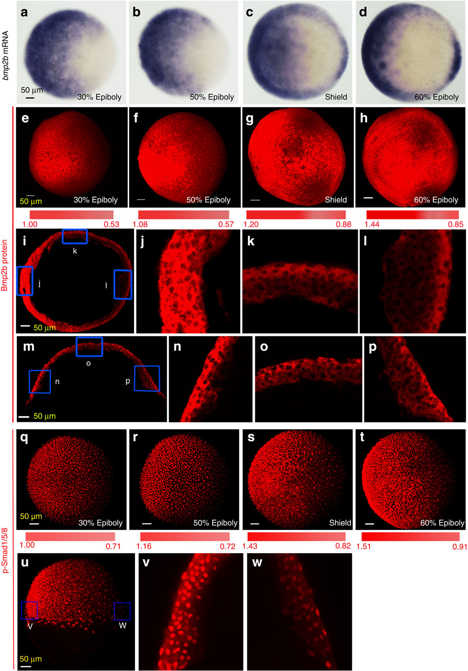
(a–d) Distribution of bmp2b transcripts revealed by whole-mount in situ hybridization at indicated stages. All embryos were animal-pole views with dorsal to the right. (e–h) Confocal microscopic detection of Bmp2b protein in whole embryos. Embryos at indicated stages were immunostained using an anti-Bmp2b antibody. The z-stack images were shown in animal-pole view with dorsal to the right. The colour bar in (e–h) illustrated signal level changes. The ratio of Bmp2b/DAPI at the two extremes was calculated and indicated. Embryos (3–5), 2–3 sections per embryos, were analysed for each group (also see Supplementary Fig. 2b, c). (i–p) Confocal microscopic detection of Bmp2b proteins in cryostat sections of embryos at the shield stage. The equatorial (i) or meridional (m) sections, which passed the organizer, were immunostained using an anti-Bmp2b antibody, and the representative z-stack images were orientated with dorsal to the right. The boxed regions in (i) and (m) were enlarged and shown in j–l and n–p, respectively. (q–t) Confocal microscopic detection of p-Smad1/5/8. Embryos were immunostained using an anti-p-Smad1/5/8 antibody. The representative z-stack images were shown in animal-pole view with dorsal to the right. (u–w) Distribution of p-Smad1/5/8 in a shield stage embryo. The embryo was laterally viewed with dorsal to the right (u) and boxed ventralmost (v) and dorsalmost (w) areas were enlarged from an optical meridional section.