
Fig. 3
- ID
- ZDB-IMAGE-140902-2
- Publication
- Al-Hamed et al., 2014 - Functional modelling of a novel mutation in BBS5
- All Figures
- Figures for Al-Hamed et al., 2014
|
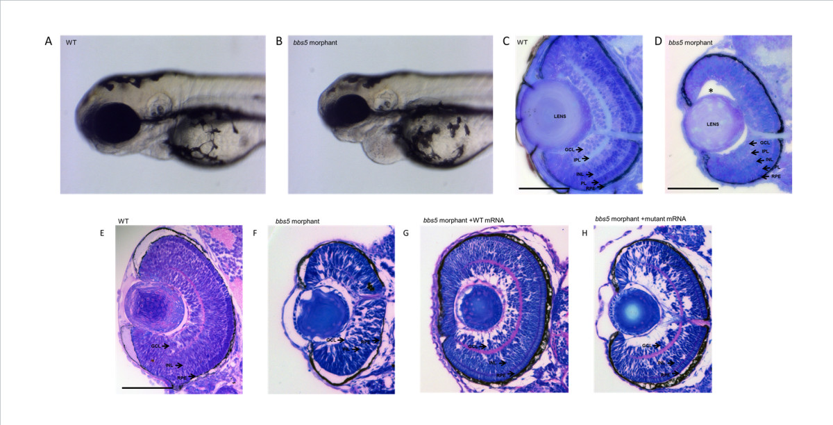
Fig. 3
Microphthalmia and retinal layering defect in bbs5 morphants and phenotypic rescue with mRNA co-injection. Light microscopy of 72 hpf embryos show a reduction in eye size in between (A) wildtype (WT) and (B)bbs5 morphants. Retinal sections demonstrating (C) WT retina with normal, well demarcated retinal layering compared to (D) morphant retina displaying partial loss of retinal layering, loss of photoreceptor layer and separation of lens from retina (*). In mRNA rescue experiments, WT mRNA or mutant mRNA was co-injected with bbs5 MO. (E) Retinal layers are preserved in WT and (F) disrupted in bbs5 morphants. (G) WT mRNA is able to rescue the retinal phenotype and eye size whilst in the (H) mutant mRNA phenotype retinal layers remain disrupted and the eye size small. Scale bar 100 um. GCL, ganglion cell layer; INL, inner nuclear layer; IPL, inner plexiform layer; PL, photoreceptor layer, RPE, retinal pigment epithelia.