
Fig. 7
- ID
- ZDB-IMAGE-140822-8
- Publication
- Whitesell et al., 2014 - An alpha-smooth muscle actin (acta2/alphasma) zebrafish transgenic line marking vascular mural cells and visceral smooth muscle cells
- All Figures
- Figures for Whitesell et al., 2014
|
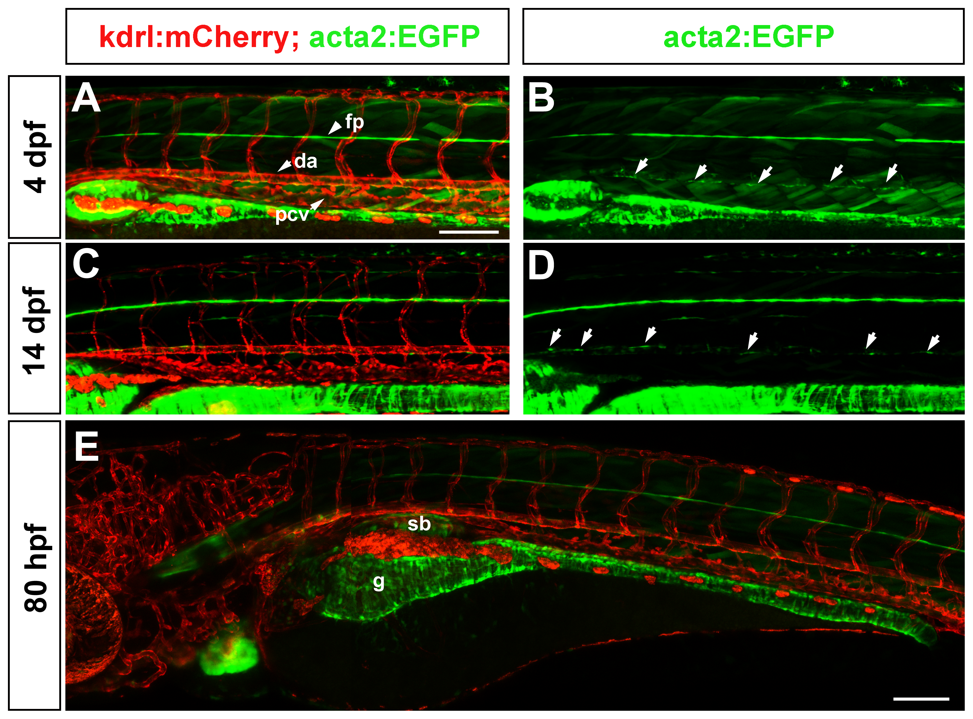
Fig. 7
Vascular and visceral mural and smooth muscle cells in the trunk.
(A–B) At 4 dpf, acta2:EGFP positive cells (arrows in B) are seen in the ventral portion of the dorsal aorta, but not in other vessels of the trunk. Floor plate (fp) expression of acta2:EGFP is observed in all images. (C–D) At 14 dpf, the distribution of vascular mural cells to the ventral portion of the dorsal aorta only, is still observed. (E) In contrast to the scarce vascular smooth muscle coverage, visceral smooth muscle cells strongly express the acta2:EGFP transgene at 80 hpf. Scale bars represent 100 μm. Green striations are skeletal muscle fibres.