
Fig. S1
- ID
- ZDB-IMAGE-140822-11
- Publication
- Whitesell et al., 2014 - An alpha-smooth muscle actin (acta2/alphasma) zebrafish transgenic line marking vascular mural cells and visceral smooth muscle cells
- All Figures
- Figures for Whitesell et al., 2014
|
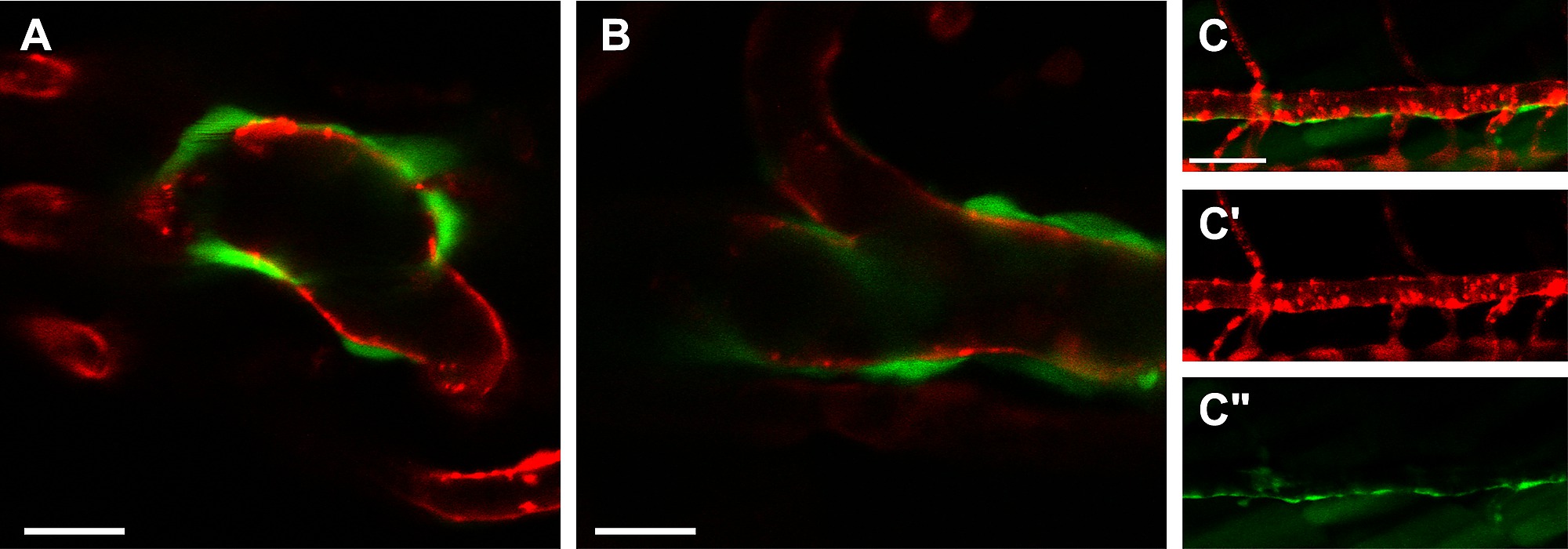
Fig. S1
The acta2:GFP transgene is expressed surrounding endothelium in the ventral aorta. Single slices of confocal micrograph stacks of double transgenic Tg(kdrl:mCherry; acta2:EGFP) zebrafish embryos at 7 dpf show green acta2:EGFP cells surrounding red kdrl:mCherry expressing endothelial cells in two different regions of the ventral aorta, distal (A) and proximate (B) to the heart outflow tract. The dorsal aorta is depicted in 11 dpf embryos, with green acta2:EGFP cells surrounding red kdrl:mCherry expressing endothelial cells (C) and with individual fluorescent markers (C′ - green acta2:EGFP cells; C′′ red kdrl:mCherry endothelial cells). Scale bar in B represents 20 μm, scale bar in C represents 50 μm.