
IMAGE
Fig. 9
- ID
- ZDB-IMAGE-140822-10
- Publication
- Whitesell et al., 2014 - An alpha-smooth muscle actin (acta2/alphasma) zebrafish transgenic line marking vascular mural cells and visceral smooth muscle cells
- All Figures
- Figures for Whitesell et al., 2014
Image
|
Figure Caption
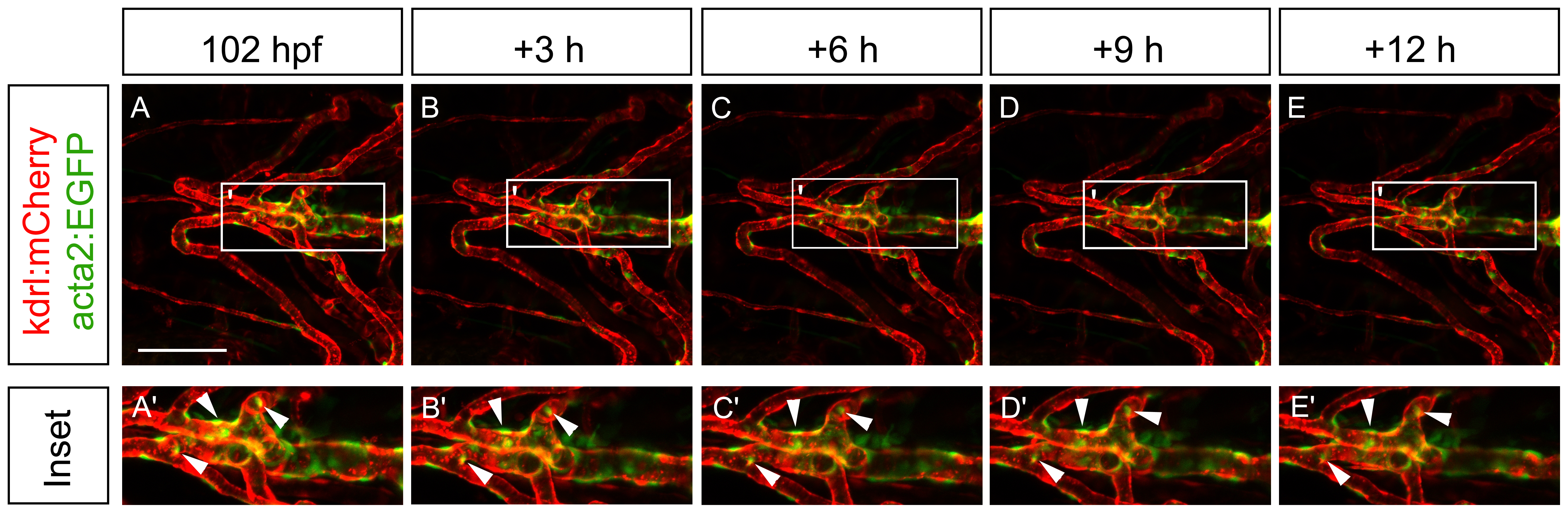
Fig. 9
Vascular mural cells of the ventral head are very stable over time.
(A-E) Single images taken from a confocal microscopy timelapse video. Images were collected at 102 hpf (A) and every three hours for 12 hours (B-E). Insets (A′-E′) show a higher magnification of the ventral aorta, where mural cells that are present at the beginning of the timelapse are still present at the end of the timelapse with no cytokinesis. Arrowheads depict mural cells throughout the timelapse that appear to have little movement. Scale bar represents 100 μm.
Acknowledgments
This image is the copyrighted work of the attributed author or publisher, and
ZFIN has permission only to display this image to its users.
Additional permissions should be obtained from the applicable author or publisher of the image.
Full text @ PLoS One