
IMAGE
Fig. S1
- ID
- ZDB-IMAGE-140811-8
- Publication
- Thiele et al., 2014 - Descending Control of Swim Posture by a Midbrain Nucleus in Zebrafish
- All Figures
- Figures for Thiele et al., 2014
Image
|
Figure Caption
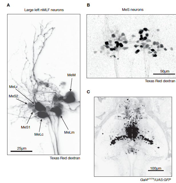
Fig. S1
Anatomy of the nMLF revealed by spinal cord backfills (related to Figure 1).
A. Confocal image projection (80 μm) of several left nMLF neurons backfilled from the spinal cord with Texas Red dextran. In this backfill, the large identified neurons MeLc, MeLr, MeLm and MeM are prominently labeled. The identity of each cell is indicated. In addition, the newly identified smaller MeS1 and MeS2 cells are indicated. B. Two-photon image projection (80 μm) of a different nMLF backfill in which many MeS neurons are labeled as defined by their small soma diameter. C. Confocal image projection (220 μm) displaying both dorsal and ventral expression patterns in Gal4s1171t/UAS:GFP.
Acknowledgments
This image is the copyrighted work of the attributed author or publisher, and
ZFIN has permission only to display this image to its users.
Additional permissions should be obtained from the applicable author or publisher of the image.
Full text @ Neuron