
|
Fig. 6
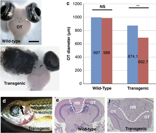
Stable transgenic fish exhibited asymmetric OT development and brain tumorigenesis. (a) A wild-type adult fish exhibited symmetric development of OT, scale bar, 1000μm. (b) A transgenic fish with gross eye tumor exhibited asymmetric OT development. (c) Comparison between left (blue bar) and right (red bar) tectal lobe diameters in the wild-type fish and between larger (blue bar) and smaller (red bar) tectal lobes in the transgenic fish showed significant difference in transgenic fish OT, but not in wild-type fish OT (n=10). NS, statistically not significant; **P<0.01. Error bars show s.d. In general, the OT from transgenic fish was smaller than wild type, because the tumor-bearing fish were smaller. (d) A 12-month-old transgenic fish showing a brain tumor (arrow); (e) hematoxylin and eosin (H&E) staining of a transverse section at the boundary of the OT and hindbrain (HB) from a wild-type fish showing symmetric OT and a small hindbrain area. (f) H&E staining of the brain tumor in (d) showing tumor formation in the HB.