
|
Fig. 3
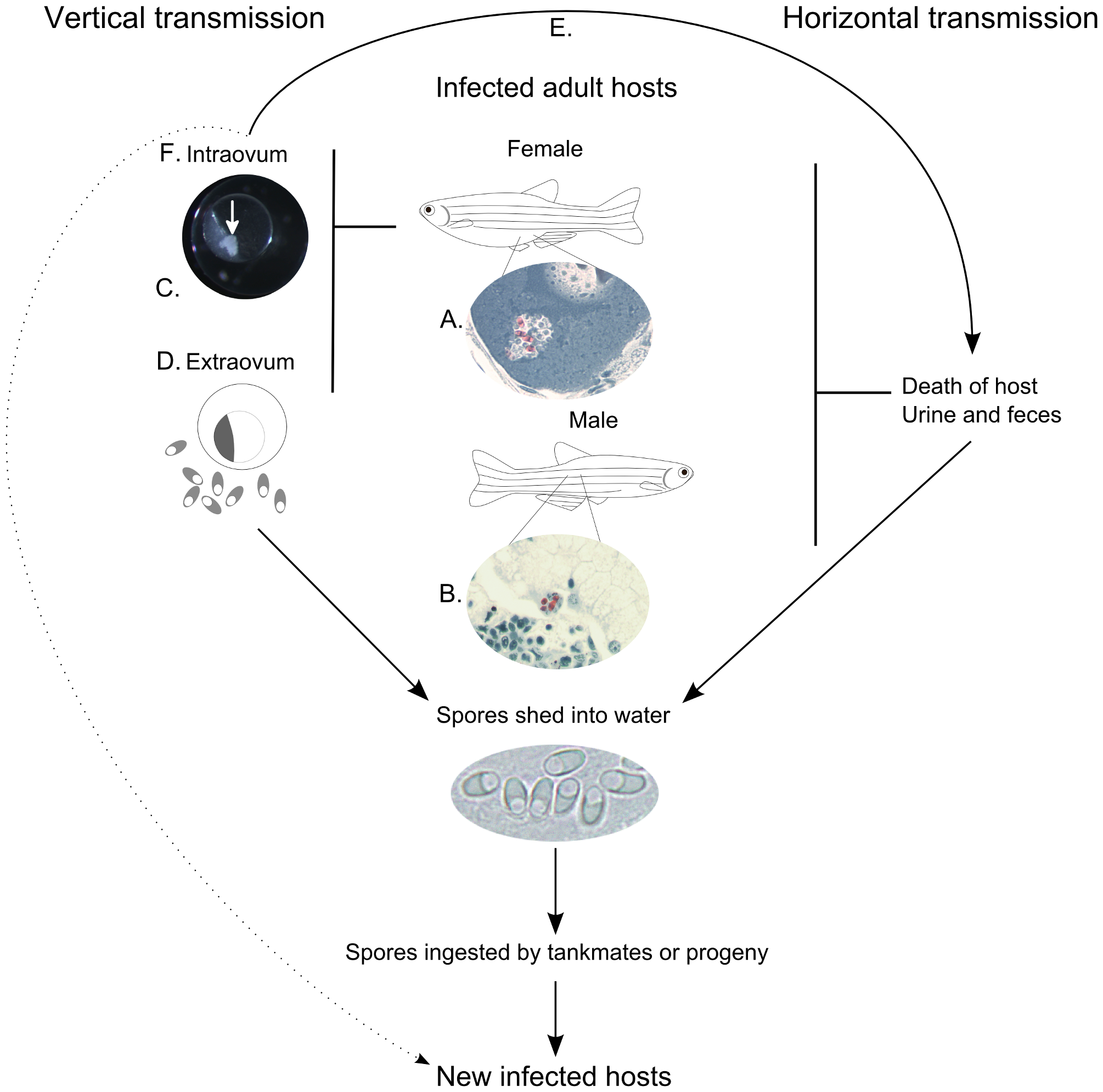
Modes of transmission of Pseudoloma neurophilia in the zebrafish, Danio rerio.
A. Luna-stained histological section showing P. neurophilia spores (red) within a secondary oocyte. Sexually mature female fish have been shown to harbor the parasite in both ovigerous stromal tissue and within various developmental stages of oocytes. B. Luna-stained histological section of kidney from an adult male zebrafish with spores present (red) within the epithelium of a renal tubule. The presence of spores in these structures is proposed to be one method by which spores can be released into the environment by live fish. C. P. neurophilia spores (arrow) present within a developing embryo. D. The presence of high numbers of P. neurophilia spores in spawn water and the high susceptibility of larval fish can result in infected progeny. E. Intraovum transmission of P. neurophilia is proposed to result in either the death of the developing embryo or larvae with the subsequent release of spores into the water infecting tank mates or F. live, infected animals that then go on to transmit the parasite horizontally and, later, vertically.