
|
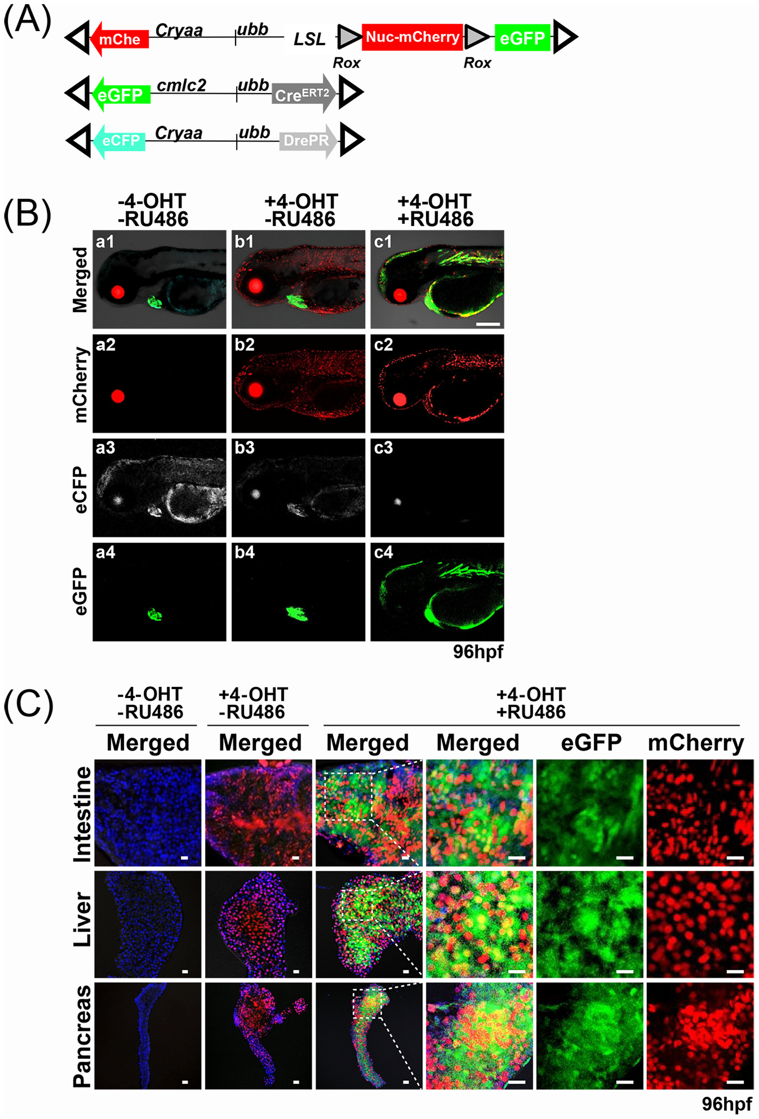
Fig. 3
Combinatorial activation of DrePR and CreERT2.
(A) Schematic of ubb:lox-stop-lox-rox-Nuc-mCherry-stop-ro x-eGFP dual reporter for assessment of both Cre- and Dre-mediated recombination, along with ubb-CreERT2 and ubb-DrePR driver lines. Ocular and cardiac fluorescence conveyed by additional cryaa:mCherry, cmlc2:eGFP and cryaa:eCFP cassettes facilitates identification of transgene-expressing embryos. Open triangles indicate Tol2 arms. (B) Triple transgenic fish were treated for 24 hrs with or without 4-OHT and RU486 as indicated, and imaged at 96 hpf. While untreated embryos showed no transgene-specific fluorescence besides that provided by the ocular mCherry, ocular eCFP and cardiac eGFP markers (Fig. 3B a1, a2, a3, and a4), embryos treated with only 4-OHT displayed widespread activation of nuc-mCherry, but no activation of eGFP (Fig. 3B b1, b2, b3, b4). In contrast, embryos simultaneously treated with both 4-OHT and RU486 displayed expression of both nuc-mCherry and eGFP (Fig. 3B c1, c2, c3, and c4). Scale bar: 200 µm. (C) Confocal imaging of dissected intestine, liver, and pancreas, confirming patterns of mCherry and eGFP expression observed in whole embryos. Following combined treatment with 4-OHT and RU486, a majority of cells in each tissue express either nuclear mCherry or cytoplasmic eGFP, with a smaller fraction of cells expressing both. Scale bar: 25 μm.