
|
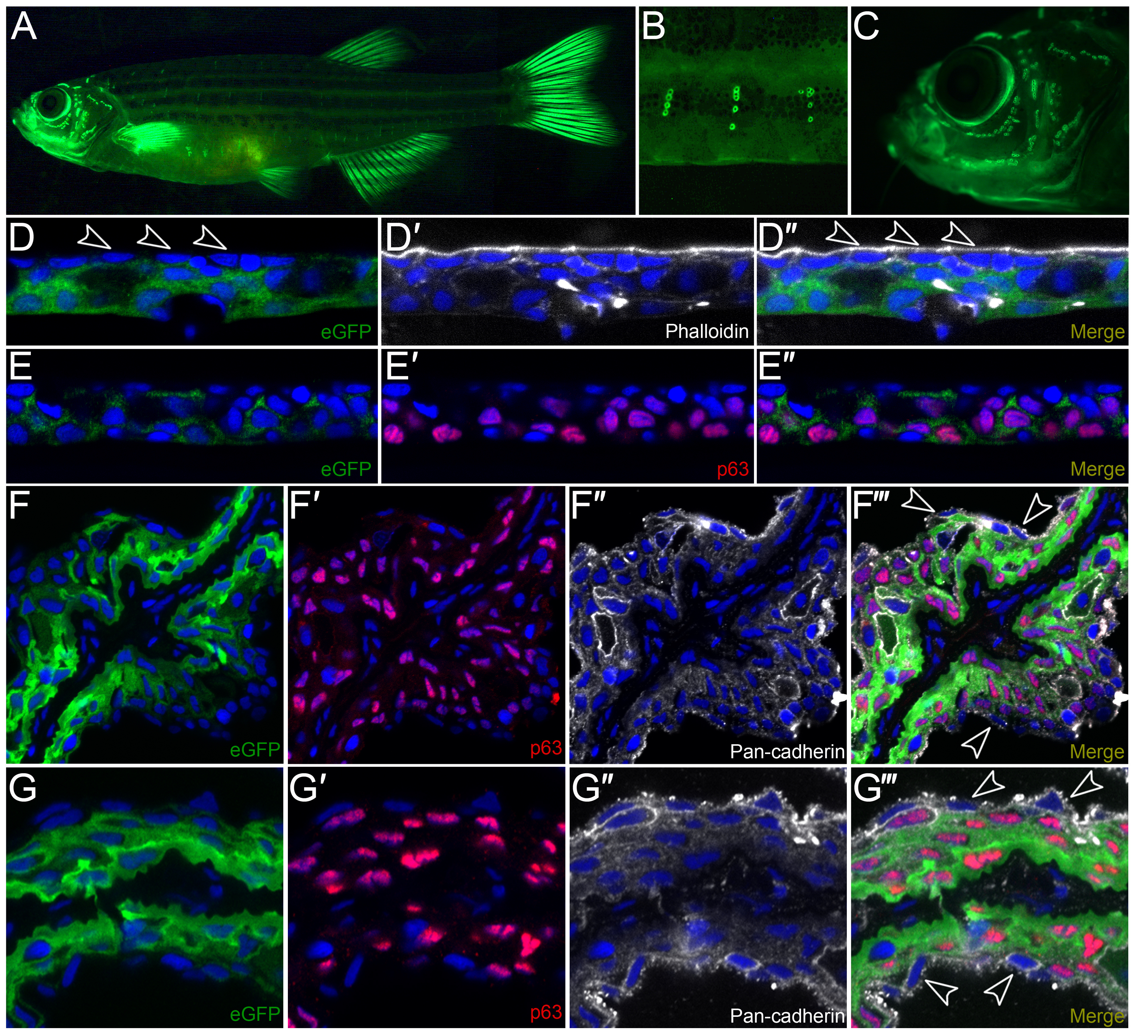
Fig. 4
Characterization of krtt1c19e:egfp transgenic adult zebrafish.
A-C: Lateral micrographs of krtt1c19e:egfp transgenic adult zebrafish, showing eGFP expression weakly in the trunk region but strongly in the fins (A). Expression is also seen associated with neuromasts of the lateral line system in the body (A, B) and head (C). D-G′′′: Immunostaining of cryosections from the trunk region (D-E′′) or fin (F-G′′′) of transgenic adults demonstrates expression is in basal and suprabasal epidermal cells, with no expression visible in the superficial epidermal stratum (arrowheads D-D′′). eGFP is shown in green (D, D′′, E, E′′, F, F′′′, G, G′′′) and co-localises with ΔNp63 (red; E′-E′′, F′, F′′′, G′, G′′′) in basal and suprabasal keratinocytes, and is excluded from the most superficial keratinocytes (e.g. arrowheads F′′′, G′′′) visualised by DAPI (blue; D-G′′′), Phalloidin staining (white, D′-D′′) or Pan-cadherin staining (white; F′′-F′′′, G′′-G′′′).