
Fig. 6
- ID
- ZDB-IMAGE-140731-38
- Genes
- Publication
- Han et al., 2014 - The Nogo-C2/Nogo receptor complex regulates the morphogenesis of zebrafish lateral line primordium through modulating the expression of dkk1b, a Wnt signal inhibitor
- All Figures
- Figures for Han et al., 2014
|
Fig. 6
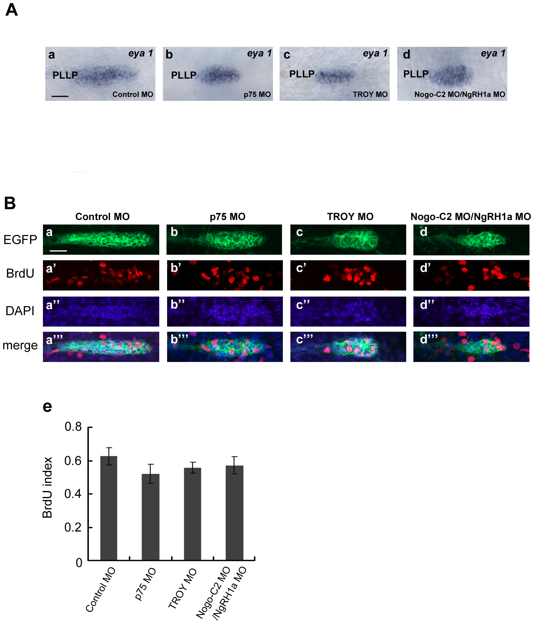
Although reduced in size, the PLL primordium of p75, TROY and Nogo-C2/NgRH1a morphants do not exhibit defects in cell proliferation.
(A) PLL primordium in control (panel a) and morphant embryos (panels b–d) were labeled by whole-mount in situ hybridization with a probe against eya1. PLLP, posterior lateral line primordium. (B) Proliferating cells in PLL primordium were examined by BrdU incorporation. Each MO was injected into the CldnB::lynEGFP transgenic line, and BrdU incorporation assays were subsequently performed from 32.5 hpf. After incorporation, the embryos were fixed and immunostained with anti-GFP and anti-BrdU antibodies. Proliferating cells in the PLL primordium are shown in control embryos (panels a–a23), and p75 (b–b23), TROY (c–c23), and Nogo-C2/NgRH1a (d–d23) morphants. Nuclei were also stained with DAPI. Scale bar, 20 μm. The BrdU index (the ratio of BrdU-positive cells/total cells) was determined in MO-injected embryos (panel e).