
Fig. 4
- ID
- ZDB-IMAGE-140730-35
- Publication
- Govindan et al., 2014 - Hapln1a is required for connexin43-dependent growth and patterning in the regenerating fin skeleton
- All Figures
- Figures for Govindan et al., 2014
|
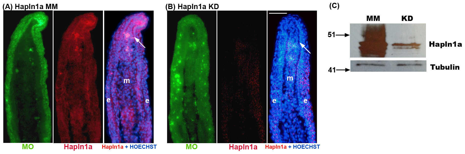
Fig. 4
Morpholino mediated knock-down of hapln1a in WT regenerating fins results in reduced Hapln1a.
Immunostaining for Hapln1a and HOECHST staining for DNA (blue). The green reveals the location of the targeting and control MOs, which are fluorescein tagged. (A) Longitudinal section of a fin ray treated with Hapln1a control morpholino (MM). (B) Longitudinal section of a fin ray knocked down for Hapln1a with a targeting morpholino (KD). Compared to the control MM fins, Hapln1a knock-down (KD) fins exhibit reduced staining for Hapln1a. (C) Immunoblots confirming reduced levels of Hapln1a protein following Hapln1a knockdown. Hapln1a-MO treated fins (KD) were compared to control morpholino (MM). Tubulin was used as a loading control. Arrow identifies the basal layer of the epidermis; m, mesenchyme; e, epithelium. Scale bar is 50 μm.