
|
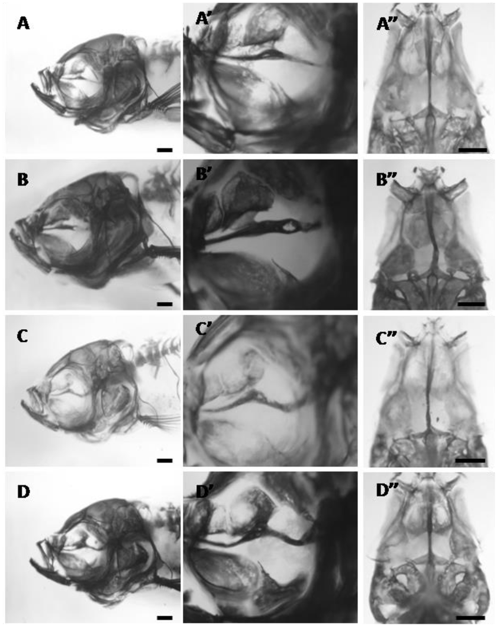
Fig. 6
The parasphenoid phenotypes in adult bone-stained zebrafish exposed to 24 hours of SMG starting at 12 hpf.
A–D) left lateral views. Higher magnifications are given in A′-D′ and in ventral view in A′′-D′′. A) slight thickening of the parasphenoid in the region where it articulates with the orbitosphenoid, 14.0 mm SL. B) thickened parasphenoid with a hole in the posterior region, 15.0 mm SL. C) parasphenoid is slightly thickened in the region where it articulates with the orbitosphenoid and severely bent posteriorly, 15.0 mm SL. D) parasphenoid is thickened at the orbitosphenoid articulation and bent towards the posterior end, 14.0 mm SL. All scale bars are 500 μm.