
Fig. 3
- ID
- ZDB-IMAGE-140730-132
- Publication
- Maradonna et al., 2013 - Probiotic Supplementation Promotes Calcification inDanio rerio Larvae: A Molecular Study
- All Figures
- Figures for Maradonna et al., 2013
|
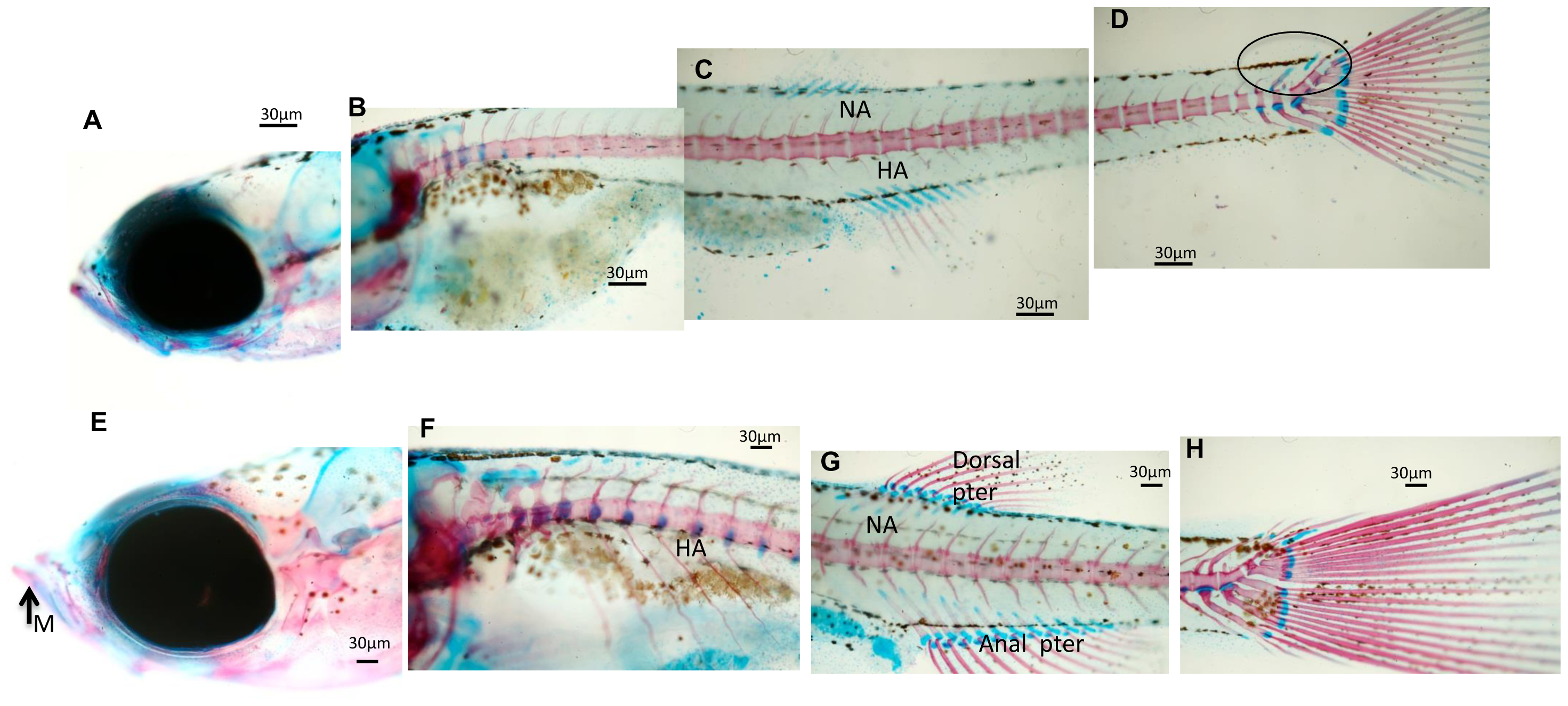
Fig. 3
Whole mount double staining of the skeleton in larvae sampled at 23 dpf.
(A–D) Images showing significant aspects of skeleton development in control zebrafish larvae. Neural arches (NA) and sketches of hemal arches (HA) are evidenced. Caudal skeleton still presents cartilaginous structures evidenced by a circle. (E–H) Representative images showing the development of the skeleton in zebrafish fed L. rhamnosus. (E) Presence of calcified mandibular (M). Neural arches (NA) and hemal arches (HA) are detected in the whole trunk of the larvae. (G) Complete formation of dorsal and anal pterygium.(H) Caudal skeleton is complete. Scale bar: 30 μm.