
Fig. 2
- ID
- ZDB-IMAGE-140730-131
- Publication
- Maradonna et al., 2013 - Probiotic Supplementation Promotes Calcification inDanio rerio Larvae: A Molecular Study
- All Figures
- Figures for Maradonna et al., 2013
|
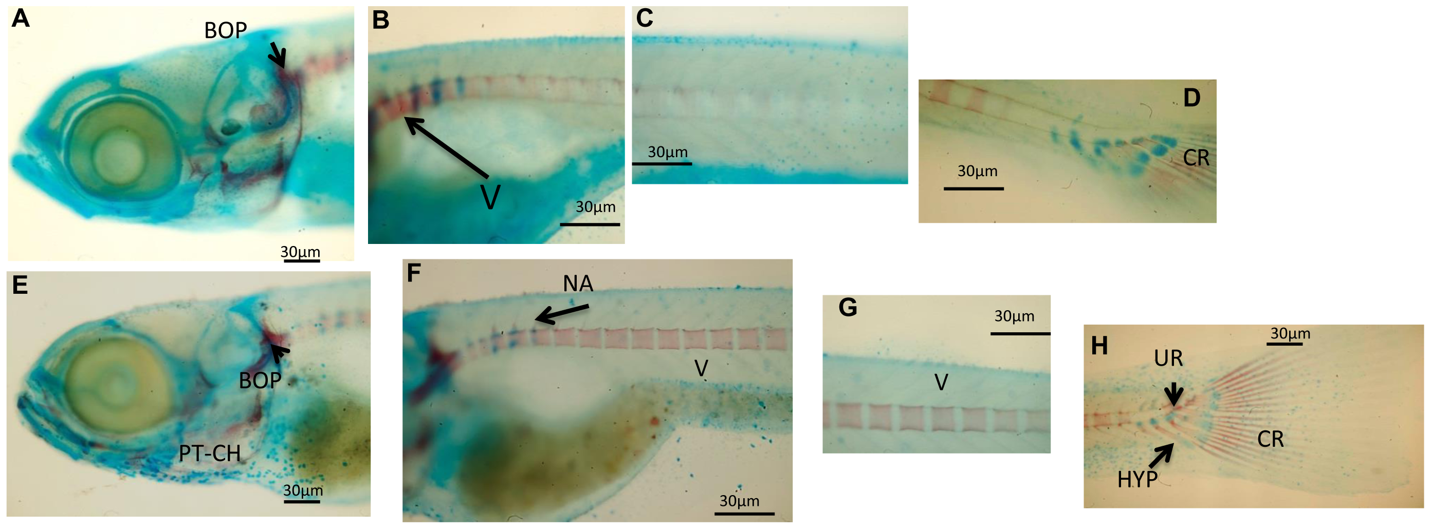
Fig. 2
Whole mounts double staining of the skeleton in larvae sampled at 16 dpf.
(A–D) Images showing significant aspects of skeleton development in control zebrafish larvae. (B) Formation of first vertebrae (V). (D) Caudal hypuralia aquires final number of structures with modified hemal arches (MHA) and caudal fin rays (CR). (E–H) representative images showing the development of the skeleton in zebrafish fed L. rhamnosus. (E) presence of calcified pharyngeal teeth (PT) and ceratohyal (CH). (F–G) Vertebrae formation (in an anterior-posterior direction) toward the posterior end of the notochord. Formation of the first neural arches (NA) is observed dorsally in the anterior vertebrae. (H) Beginning of calcification of the hypurals (HYP) under the urostyle (UR) and presence of calcified caudal fin rays. Scale bar: 30 μm.