
Fig. 1
- ID
- ZDB-IMAGE-140730-130
- Publication
- Maradonna et al., 2013 - Probiotic Supplementation Promotes Calcification inDanio rerio Larvae: A Molecular Study
- All Figures
- Figures for Maradonna et al., 2013
|
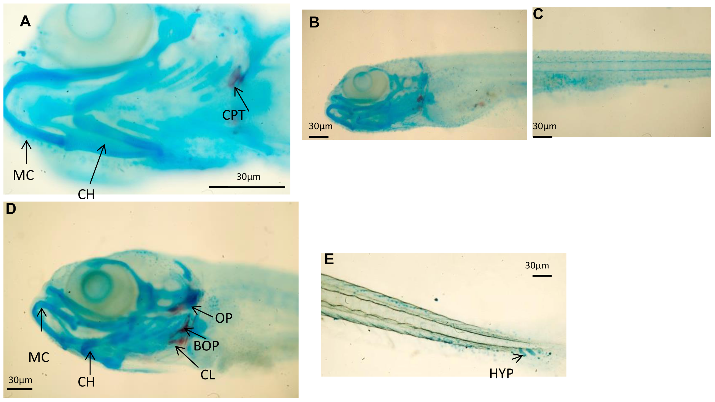
Fig. 1
Skeletal development in zebrafish using alcian blue-Alizarin red double staining.
A 9 dpf zebrafish control larvae head skeleton presenting calcified calcified pharyngeal teeth (CPT)while other structures like Meckel′s cartilage (MC) and ceratohyal (CH) remain as cartilage); (B–C) 9 dpf control zebrafish head (B) and trunk (C) presenting no signals of bone calcification.(A) A 9 dpf L. rhamnosus fed zebrafish larvae head skeleton presenting calcification of the opercula (OP), cleithrum (CL) and basioccipital articulatory process (BOP). Meckel′s cartilage (MC) and ceratohyal (CH) remain as cartilage; (E) 9 dpf L. rhamnosus fed zebrafish presenting the first hypurals (HYP) developing. Scale bar: 30 μm.