
Image
|
Figure Caption
Fig. 7
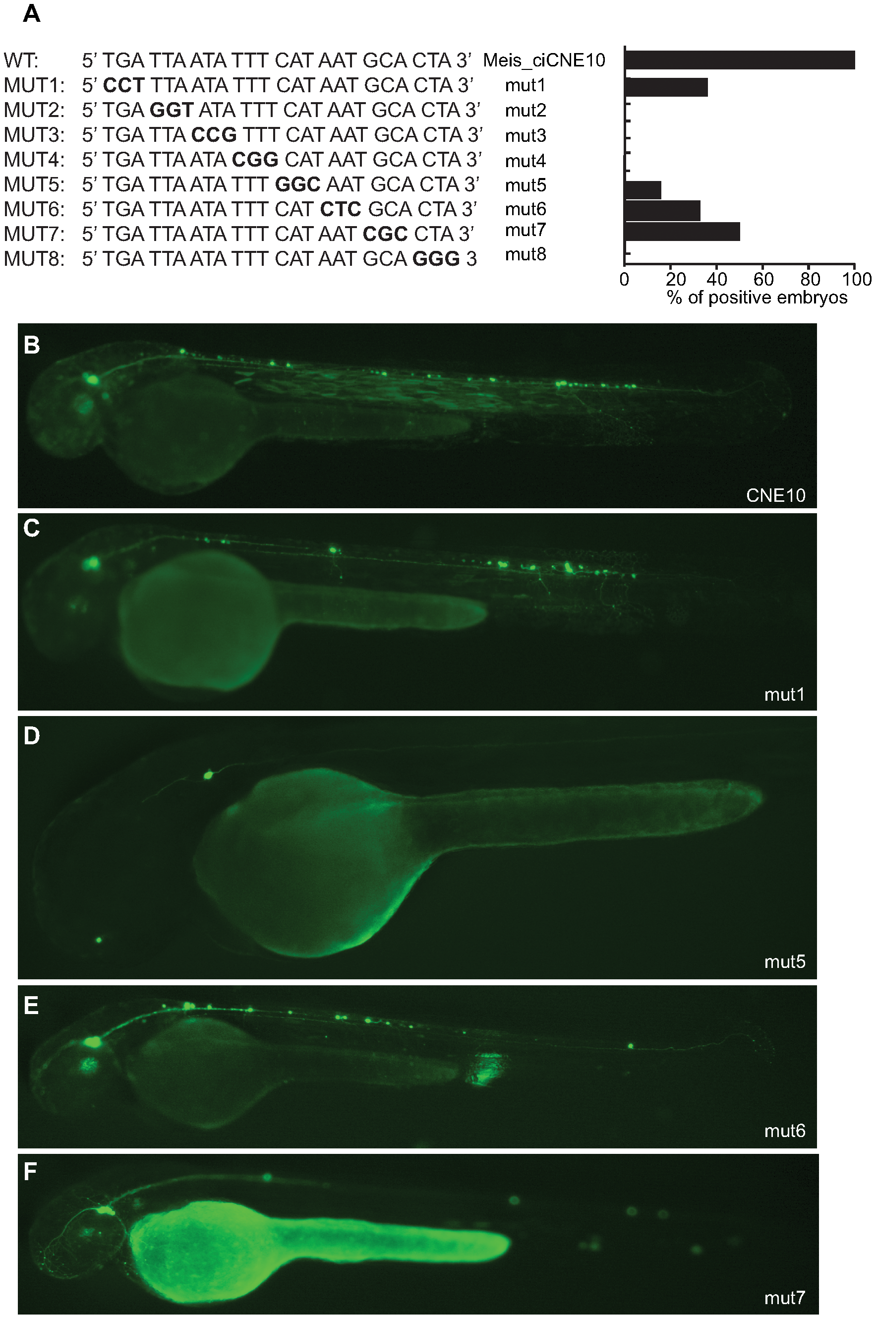
Mutational analysis of putative binding site in CNE10.
(A) Schematic representation and quantification of mutations introduced in the 24 nt core sequence of CNE10. Mutated triplets are shown in bold. (B) Embryos injected with wt CNE10 show GFP expression in the nervous system at 48 hpf. (C, E) Embryos injected with mut1 and mut 6 show a GFP expression pattern similar to the wt construct, whereas injection of mut5 (D) drives GFP expression only in few neuronal cells. (F) In embryos injected with mut7 GFP expression is mainly detected in trigeminal ganglion neurons.
Acknowledgments
This image is the copyrighted work of the attributed author or publisher, and
ZFIN has permission only to display this image to its users.
Additional permissions should be obtained from the applicable author or publisher of the image.
Full text @ PLoS Genet.