
IMAGE
Fig. S2
- ID
- ZDB-IMAGE-140728-11
- Publication
- Asaoka et al., 2014 - The Hippo Pathway Controls a Switch between Retinal Progenitor Cell Proliferation and Photoreceptor Cell Differentiation in Zebrafish
- All Figures
- Figures for Asaoka et al., 2014
Image
|
Figure Caption
Fig. S2
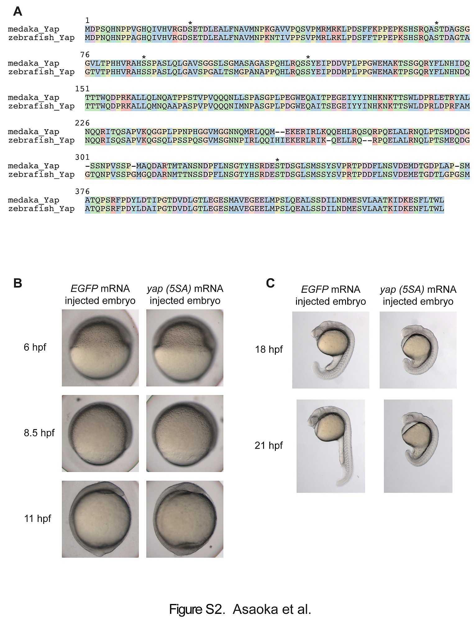
Morphological analysis of yap (5SA) mRNA-injected zebrafish embryos during the gastrulation and segmentation periods. (A) Alignment of amino acid sequence of medaka Yap with its zebrafish homolog performed as in Fig. S1. *, conserved serine residues phosphorylated by Lats. (B) Representative images of yap (5SA) mRNA-injected zebrafish embryos (N = 3) at the indicated developmental stages during gastrulation. Embryos were injected with EGFP mRNA as a control. (C) Representative lateral images of the embryos in (B) examined at the indicated stages during segmentation.
Acknowledgments
This image is the copyrighted work of the attributed author or publisher, and
ZFIN has permission only to display this image to its users.
Additional permissions should be obtained from the applicable author or publisher of the image.
Full text @ PLoS One