
Fig. 4
- ID
- ZDB-IMAGE-140724-11
- Publication
- Clay et al., 2014 - Cadherin 6 promotes neural crest cell detachment via F-actin regulation and influences active Rho distribution during epithelial-to-mesenchymal transition
- All Figures
- Figures for Clay et al., 2014
|
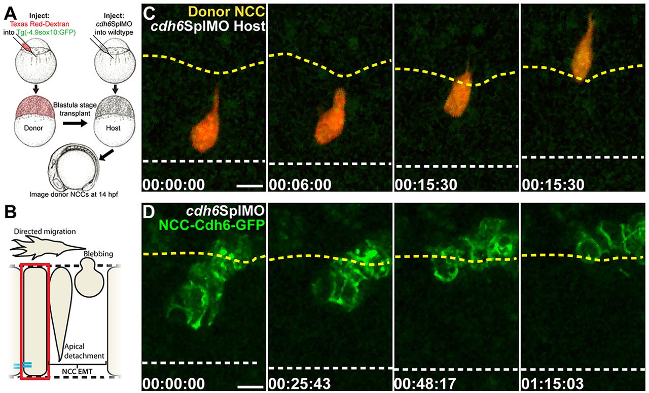
Fig. 4
Cdh6 function in EMT is NCC autonomous and Cdh6-GFP rescues morpholino knockdown. (A) Cell transplantation experiments. (B) Imaging region in C,D. (C,D) Confocal z-projections (dorsal views, anterior left) showing NCCs that were in the neuroepithelium at imaging onset. Time-lapse imaging began at 14hpf. Yellow dashed lines mark basal neuroepithelial surfaces and white dashed lines mark apical midlines. (C) A wild-type donor NCC (red/green) undergoes EMT normally when transplanted into a Cdh6 knockdown embryo. (D) NCCs expressing Cdh6-GFP undergo EMT as a cluster in Cdh6 knockdown embryo. Time=h:min:s. Scale bars: 10μm.