
IMAGE
Fig. 7
- ID
- ZDB-IMAGE-140715-75
- Genes
- Publication
- Mackereth et al., 2005 - Zebrafish pax8 is required for otic placode induction and plays a redundant role with Pax2 genes in the maintenance of the otic placode
- All Figures
- Figures for Mackereth et al., 2005
Image
|
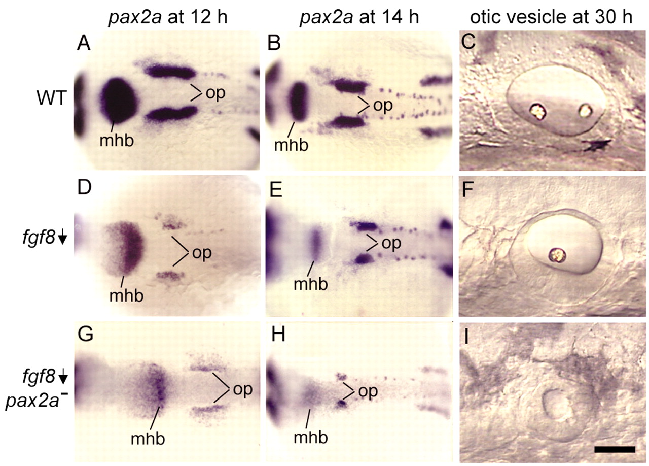
Figure Caption
Fig. 7 pax2a interacts with fgf8. Wild-type embryos (A-C), ace (fgf8) single mutants (D-F) and ace-noi (fgf8-pax2a) double mutants (G-I). Images show dorsal views of pax2a expression at 12 hpf (A,D,G) and 14 hpf (B,E,H), and lateral views of otic vesicles at 30 hpf (C,F,I). The specimen in B is the same as in Fig. 5A, and the specimen in C is the same as in Fig. 2B. Anterior is to the left in all specimens. mhb, midbrain-hindbrain border; op, otic placode. Scale bar in I: 170 μm for A,B,D,E,G,H; 35 μm for C,F,I.
Figure Data
Acknowledgments
This image is the copyrighted work of the attributed author or publisher, and
ZFIN has permission only to display this image to its users.
Additional permissions should be obtained from the applicable author or publisher of the image.
Full text @ Development