
Fig. 6
- ID
- ZDB-IMAGE-140715-74
- Publication
- Mackereth et al., 2005 - Zebrafish pax8 is required for otic placode induction and plays a redundant role with Pax2 genes in the maintenance of the otic placode
- All Figures
- Figures for Mackereth et al., 2005
|
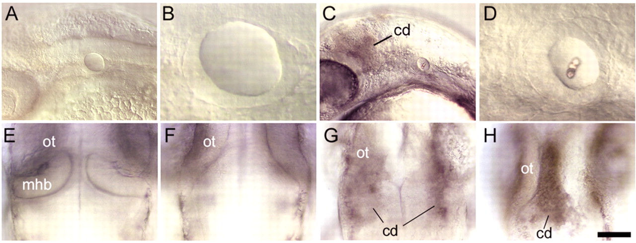
Fig. 6 Distinct functions of pax8 splice isoforms. (A-D) Lateral views of the head and otic structures at 30 hpf in noi (pax2a) mutants injected either with pax8 variant 1 MO (A,B) or noi mutants injected with pax8 variant 2/3 MO (C,D). Most noi mutants knocked down for variant 1 isoforms produce a moderate-sized otic vesicle containing hair cells but lacking otoliths (B). In contrast, noi mutants knocked down for variant 2 and 3 isoforms typically produce a small otic vesicle containing both hair cells and otoliths (D) or no otic vesicle at all (data not shown). In addition, all noi mutants knocked down for variant 2 and 3 isoforms show persistent cell death (cd) in the midbrain-hindbrain region. (E-H) Dorsal views of the midbrain-hindbrain border region at 30 hpf in an uninjected wild-type embryo (E), an uninjected noi mutant (F), a noi mutant injected with pax8 variant 1 MO (G) and a noi mutant injected with pax8 variant 2/3 MO (H). Increased cell death is not evident in the majority of noi mutants knocked down for variant 1 isoforms (A) and, if present (G), cell death is diffuse and limited to dorsolateral tissue. In noi mutants knocked down for variant 2 and 3 isoforms, cell death is invariably present, intense, and localized to the midline of the midbrain-hindbrain border region (H). Anterior is to the left (A-D) or to the top (E-F). cd, cell death; mhb, midbrain-hindbrain border; ot, optic tectum. Scale bar in H: 75 μm for A,C; 50 μm for E-H; 19 μm for B,D.