
Fig. S1
- ID
- ZDB-IMAGE-140710-48
- Genes
- Publication
- Bu et al., 2014 - Protein tyrosine phosphatase PTPN9 regulates erythroid cell development through STAT3 dephosphorylation in zebrafish
- All Figures
- Figures for Bu et al., 2014
|
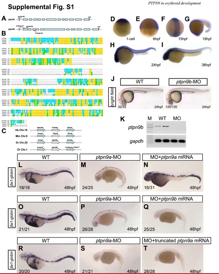
Fig. S1 Ptpn9a, but not ptpn9b, is crucial for early development in zebrafish. (A) Genomic structure of ptpn9a and ptpn9b in zebrafish, showing morpholino targeting sites (ptpn9a-MO and ptpn9b-MO) and RT-PCR primers for Fig. S2H by the red lines. (B) Protein sequence alignments among human PTPN9 (hPTPN9), mouse Ptpn9 (mPtpn9), and zebrafish Ptpn9a (ptpn9a) and Ptpn9b (ptpn9b). Note hPTPN9, mPtpn9 and Ptpn9a are highly conserved while Ptpn9b has unique linker amino-acids. (C) Genomic synteny shows that Ptpn9a is a potential ortholog of hPTPN9 and mPtpn9, while Ptpn9b was derived from zebrafish genome duplication. (D-I) RNA in situ hybridization was performed with a ptpn9b probe in 1-cell (D), 6 hpf (E), 15 hpf (F), 18 hpf (G), 24 hpf (H), and 36 hpf (I) embryos. (J-K) No apparent blood defects were found in ptpn9b morphants (J, right panel), compared with wild-type controls (J, left panel). Ptpn9b-MO was injected at 10 ng per embryo. Semi-quantitative RT-PCR revealed that ptpn9b was not detectable in ptpn9b morphants (K), suggesting that ptpn9b-MO was effective. Gapdh was used as an internal control. (L-N) Co-injection with synthetic zebrafish ptpn9a mRNA rescued βe1-globin and embryo morphology in ptpn9a morphant embryos at 48 hpf (N), compared with controls (L). To prevent binding of synthetic zebrafish ptpn9a mRNA to ptpn9a-MO, we added the kozak sequence (GCCACC) before the coding district sequence (CDS) of ptpn9a. (O-T) Over-expression of either ptpn9b mRNA or truncated ptpn9a mRNA that the SEC14 domain of Ptpn9a was deleted had no rescue on βe1-globin and embryo morphology in ptpn9a morphants at 48 hpf (Q, T), compared with controls (O, R). Lateral views are shown with anterior to the left (G-J, L-T), dorsal views are shown with anterior to the top (F).