
|
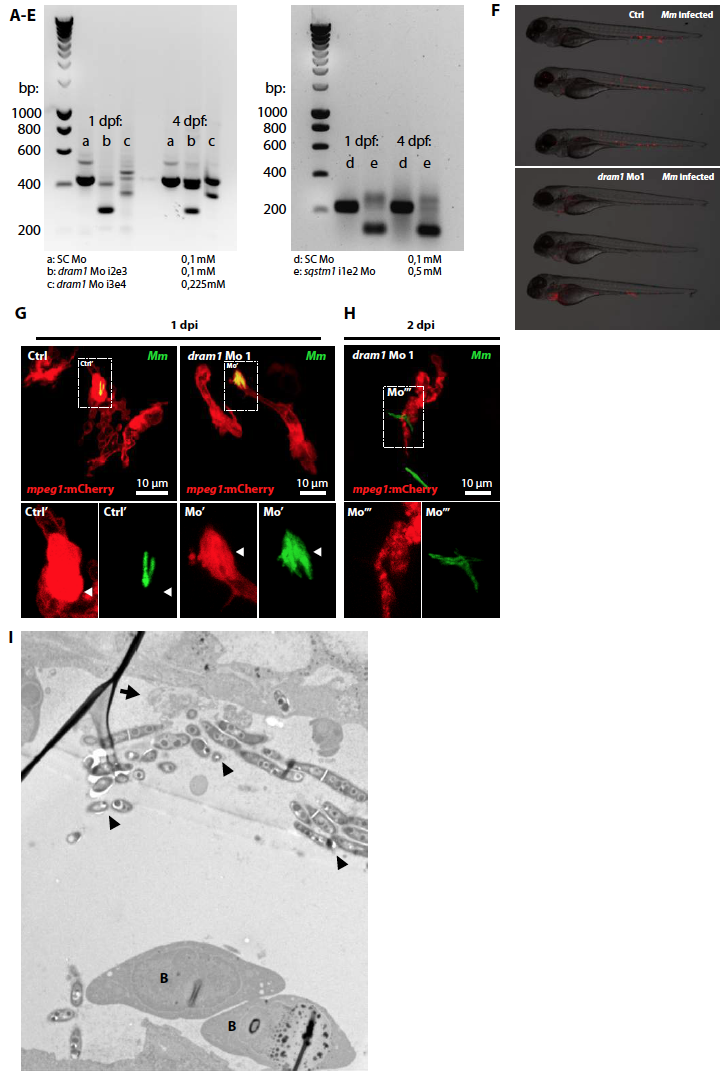
Fig. S5
Dram1 knockdown effect in zebrafish embryos.
Reverse transcription polymerase chain reaction (RT-PCR) was used to confirm antisense morpholino blocking of intron-exon splicing events in zebrafish dram1 or sqstm1 at 1 and 4 days post fertilization. (A) and (D) 0,1 mM Standard control (SC) morpholino; (B) 0,1 mM dram1 Mo 1 (i2e3, targeting the splicing event between the second intron and the third exon); (C) 0,225 mM dram1 Mo 2 (i3e4, targeting the splicing event between the third intron and the fourth exon); (E) 0,5 mM sqstm1 Mo (i1e2, targeting the splicing event between the second intron and the third exon). For both dram1 and sqstm1, the RT-PCR product (approximately 400 and 200 bp in size, respectively) is disrupted upon morpholino injection. Morpholino sequences and primers used for the RT-PCR are listed in supplementary tables 1 and 2. Supplementary figure 5, related to figure 5. Dram1 knockdown effect in zebrafish embryos
(F) figure 5. Equal bacterial burdens in control and dram1 morpholino injected embryos. Representative stereo micrographs of standard control or dram1 morpholino injected embryos. Control injected embryos were infected with 200 CFU Mm versus 50 CFU for dram1 morphants to obtain equal bacterial burdens at the time point of RNA isolation.
(G-H) Mycobacterial localization in or outside of macrophages. Representative confocal micrographs of mpeg1:mCherry transgenic embryos injected with standard control or dram1 morpholino 1 and infected with GFP-labeled Mm (A: 1 dpi; B: 2 dpi). Boxed areas are detailed below with the green and red channel shown separately. Arrowheads indicate bacteria enclosed by membranes.
(I) Transmission electron micrograph of a dram1 morpholino injected embryo infected with Mm. Clusters of extracellular bacteria (arrowheads) are residing inside a blood vessel, as illustrated by the presence of red blood cells (B). The remnants of a ruptured cell are indicated by an arrow.