
|
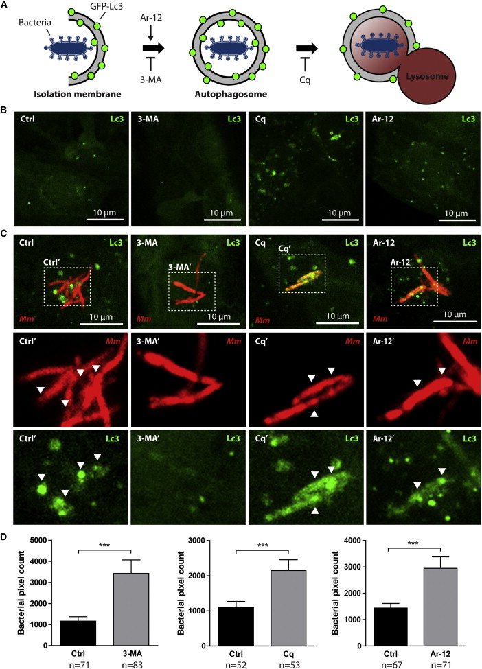
Fig. 1
Stress-Induced Autophagy Is Not Beneficial for Defense of Zebrafish Embryos against Mycobacterial Infection
(A) Schematic representation of the effects of 3-MA, Cq, and Ar-12 on autophagosome formation and autophagic flux.
(B) GFP-Lc3 embryos 2 dpf were treated for 24 hr with DMSO (control), 3-MA, Cq, or Ar-12. Representative confocal micrographs of endothelial cells at 3 dpf are shown.
(C) GFP-Lc3 embryos treated as described for (A) injected with Mm. Representative confocal micrographs of infected cells at 3 dpf are shown. Boxed areas are detailed below; arrowheads indicate overlap between Mm and Lc3.
(D) AB/Tupfel long fin (AB/TL) embryos treated as described for (A) infected with Mm. Bacterial pixel counts were determined at 3 dpi.
Data (mean ± SEM) is pooled from at least two individual experiments (n e 50 embryos per group). See also Figure S1.