
Fig. 7
- ID
- ZDB-IMAGE-140702-10
- Publication
- van der Vaart et al., 2014 - The DNA Damage-Regulated Autophagy Modulator DRAM1 Links Mycobacterial Recognition via TLR-MYD88 to Autophagic Defense
- All Figures
- Figures for van der Vaart et al., 2014
|
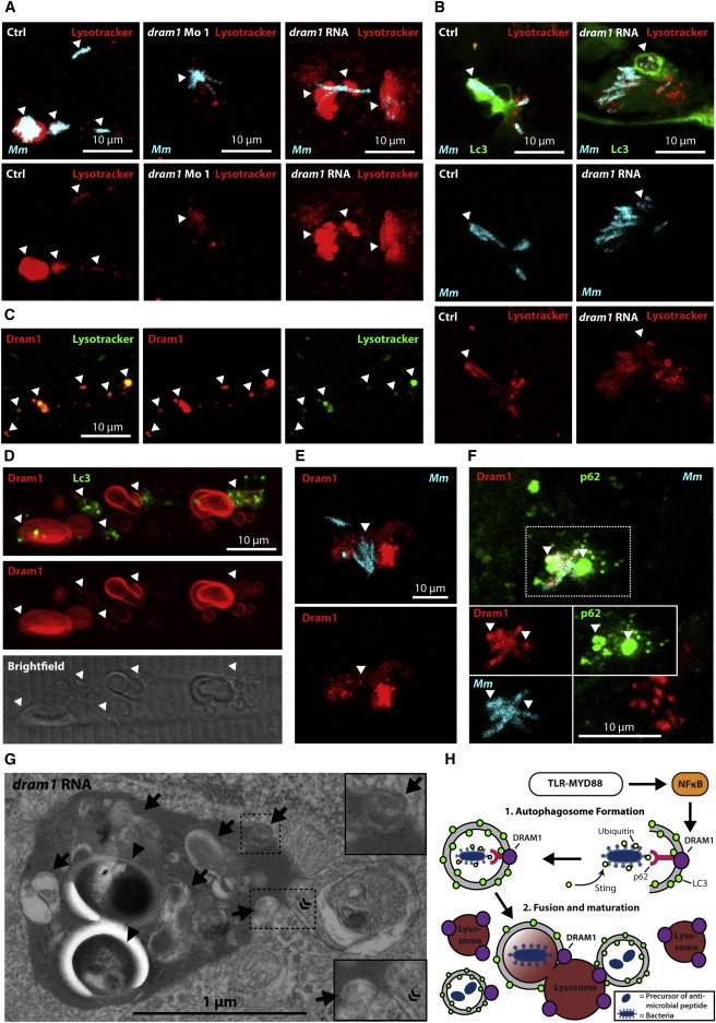
Fig. 7
Dram1 Mediates Autophagic Flux and Lysosomal Maturation via Multiple Vesicle Fusion Events
(A and B) Embryos were infected with crimson-labeled Mm and stained with LysoTracker Red (arrowheads indicate colocalization). (A) Wild-type embryos injected with standard control, dram1 morpholino, or dram1 RNA (100 pg). (B) dram1 RNA- or control-injected GFP-Lc3 embryos.
(C–F) Embryos transiently expressing mCherry-Dram1, colocalized with (C) LysoTracker Green, (D) GFP-Lc3, (E) crimson-labeled Mm, or (F) crimson-labeled Mm, and immunohistochemistry detection of p62 (arrowheads indicate colocalization).
(G) Transmission electron micrograph of dram1 RNA-injected embryos infected with Mm (arrowheads). Arrows indicate (remnants of) vesicle fusion, and « indicates the double membrane of an autophagosome.
(H) Schematic representation of the findings presented in this manuscript, as explained in the Discussion. See also Figure S7.