
Fig. S9
- ID
- ZDB-IMAGE-140626-12
- Publication
- Okigawa et al., 2014 - Different combinations of Notch ligands and receptors regulate V2 interneuron progenitor proliferation and V2a/V2b cell fate determination
- All Figures
- Figures for Okigawa et al., 2014
|
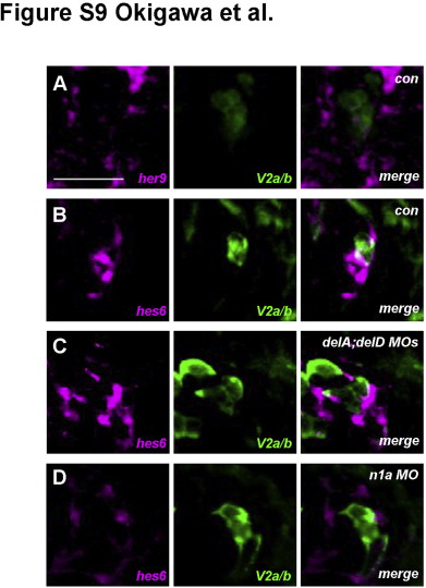
Fig. S9 Expression patterns of her9 and hes6 in and around V2 neuronal lineage cells. her9 mRNA Expression was detected in the spinal cord but not within V2 neurons in control embryos (A). In contrast, slightly higher hes6 mRNA expression was observed in one of a pair of V2 neurons (B). In deltaA/deltaD double MO-injected embryos, some but not all of the GFP-positive V2 cells (V2a or V2b) expressed hes6 (C). In notch1a knockdown embryos, GFP-positive V2 cells (most of which were V2a and not V2b) expressed her6 very weakly (D). Transverse sections through the trunk region in embryos at 24 hpf. All images are of the right lower portion of the neural tube. The mRNAs were mainly in the cytoplasm, whereas GFP was in both the nucleus and cytoplasm. Bar: 20 µm.
Reprinted from Developmental Biology, 391(2), Okigawa, S., Mizoguchi, T., Okano, M., Tanaka, H., Isoda, M., Jiang, Y.J., Suster, M., Higashijima, S.I., Kawakami, K., Itoh, M., Different combinations of Notch ligands and receptors regulate V2 interneuron progenitor proliferation and V2a/V2b cell fate determination, 196-206, Copyright (2014) with permission from Elsevier. Full text @ Dev. Biol.