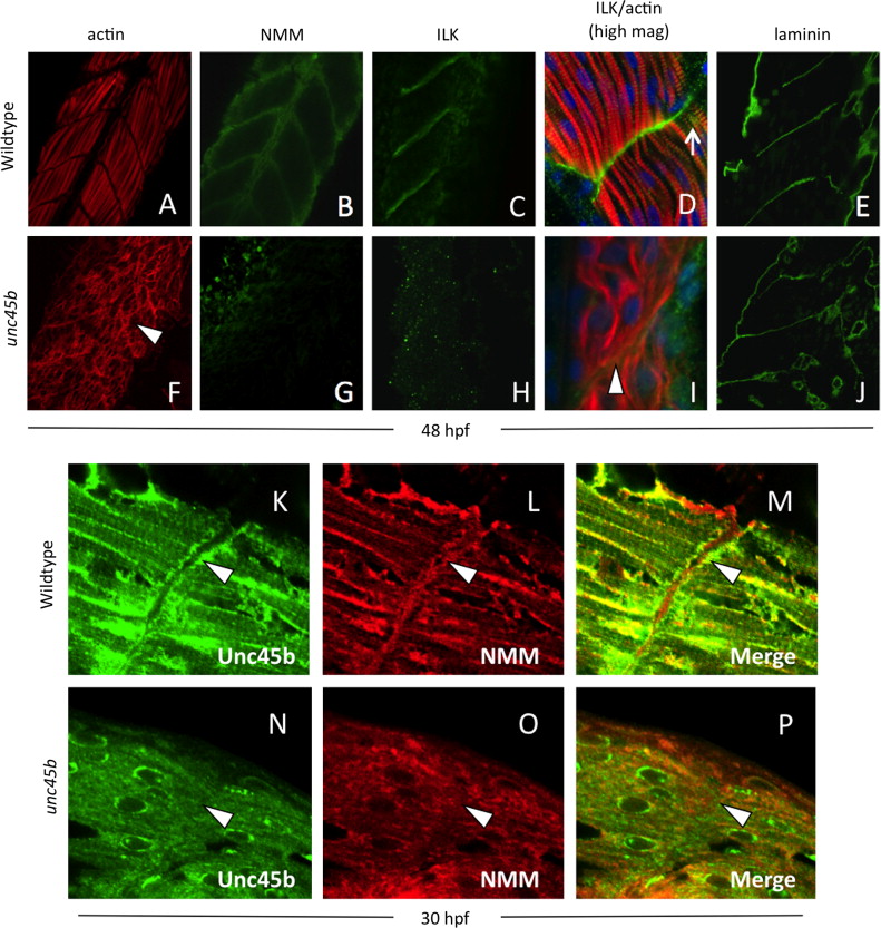
Fig. 8 Localization of Unc45b, NMM and costamere components to the myoseptum is lost in unc45b mutant embryos. Immunofluorescent staining of WT (A–E) and unc45b mutant embryos (F–J) at 24+ hours post-fertilization. The positions of myofiber attachment points at the myosepta were made visible by actin staining (A, F). Like early embryos, mature muscle tissue was characterized by the localization of non-muscle myosin (B) and integrin-linked kinase (C, D) to the myosepta in WT embryos. This localization was lost in unc45b mutants (G, H, I). At higher magnification (D, I), ILK co-localizes with actin striations at lateral costamere attachment points (arrow in D); this pattern of staining was also lost in unc45b mutant embryos (I). Green fluorescence to the right of the actin staining represents background staining outside of the muscle tissue, and blue fluorescence indicates DAPI counter-staining. However, the tissue morphology was not changed in mutants, and the disorganized myofibers were still separated by myosepta (arrowhead in I) containing extracellular matrix components such laminin (compare panels E and J). Furthermore, enrichment of Unc45b (K) and NMM (L) at the myosepta in older embryos (arrowheads) was lost in unc45b mutants (N, O), and co-localization of these proteins was greatly reduced (compare M and P).
Reprinted from Developmental Biology, 390, Myhre, J.L., Hills, J.A., Jean, F., Pilgrim, D.B., Unc45b is essential for early myofibrillogenesis and costamere formation in zebrafish, 26-40, Copyright (2014) with permission from Elsevier. Full text @ Dev. Biol.

