
IMAGE
Fig. S1
- ID
- ZDB-IMAGE-140613-21
- Publication
- Dickover et al., 2014 - The atypical Rho GTPase, RhoU, regulates cell-adhesion molecules during cardiac morphogenesis
- All Figures
- Figures for Dickover et al., 2014
Image
|
Figure Caption
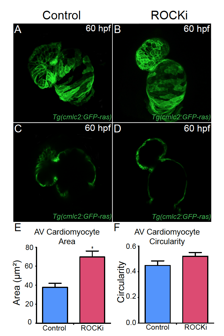
Fig. S1 ROCK inhibitor treated hearts exhibit AV canal defects. (A, B) Confocal projections and (C, D) cross-sectional analyses of 60 hpf Tg(cmlc2:ras-eGFP) hearts reveal that (B, D) ROCK inhibitor (ROCKi) treated zebrafish exhibit cardiac looping and AV canal defects. (E, F) Bar graphs represent cardiomyocyte (E) surface area and (F) morphology/circularity measurements of Tg(cmlc2:ras-eGFP) hearts at 60 hpf. Mean+s.e.m. Studentós t-test, *p<0.05 (n = 12 for control DMSO-treated hearts and n = 12 for ROCK inhibitor treated hearts).
Acknowledgments
This image is the copyrighted work of the attributed author or publisher, and
ZFIN has permission only to display this image to its users.
Additional permissions should be obtained from the applicable author or publisher of the image.
Reprinted from Developmental Biology, 389, Dickover, M., Hegarty, J.M., Ly, K., Lopez, D., Yang, H., Zhang, R., Tedeschi, N., Hsiai, T.K., Chi, N.C., The atypical Rho GTPase, RhoU, regulates cell-adhesion molecules during cardiac morphogenesis, 182-91, Copyright (2014) with permission from Elsevier. Full text @ Dev. Biol.