
Fig. 4
- ID
- ZDB-IMAGE-140613-17
- Genes
- Antibodies
- Publication
- Dickover et al., 2014 - The atypical Rho GTPase, RhoU, regulates cell-adhesion molecules during cardiac morphogenesis
- All Figures
- Figures for Dickover et al., 2014
|
Fig. 4
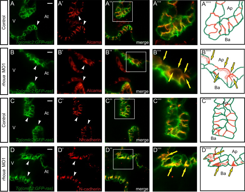
The cell adhesion proteins in the AV cardiomyocytes of rhoua morpholino knockdown zebrafish hearts are mis-localized. Confocal imaging of Tg(cmlc2:ras-eGFP) control and rhoua ATG MO1 knockdown zebrafish hearts that were examined by (A, B) Alcama antibody and (C, D) N-Cadherin antibody at 60 hpf reveals mislocalization of Alcama and N-cadherin in rhoua MO1 knockdown zebrafish AV cardiomyocytes. (A-D) Tg(cmlc2:ras-eGFP)s883 AV cardiomyocytes, (A′-D′) Immunohistochemical staining of AV cardiomyocytes, (A′′-D′′) Merge of Tg(cmlc2:ras-eGFP)s883 and immunohistochemical staining analysis, (A′′′-D′′′) Boxed area of A′′-D′′, and (A′′′′-D′′′′) Schematic representation of A′′′-D′′′. White arrowhead - AV cardiomyocytes, yellow arrows - mislocalized Alcama and N-cadherin immunostaining. At - atrium, V - ventricle, Ap - apical, Ba - basal. White scale bar - 10 µM.
Reprinted from Developmental Biology, 389, Dickover, M., Hegarty, J.M., Ly, K., Lopez, D., Yang, H., Zhang, R., Tedeschi, N., Hsiai, T.K., Chi, N.C., The atypical Rho GTPase, RhoU, regulates cell-adhesion molecules during cardiac morphogenesis, 182-91, Copyright (2014) with permission from Elsevier. Full text @ Dev. Biol.