
|
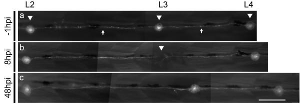
Fig. 3
Ablation and regeneration of a single posterior lateral line neuromast. (a-c)Tg(cxcr4b:mRFP) fish show labeling of posterior lateral line neuromasts and interneuromastic cells. (a) Lateral view of 72 hpf control larva showing intact lateral line neuromasts (arrowheads) connected by interneuromastic cells (arrows). (b) Two 8 μA pulses of 2 seconds each were applied to the L3 posterior lateral line neuromast. Complete dissapearance of the neuromast’s cells is observed (arrowhead) while adjacent neuromasts (L2 and L4) and interneuromastic cells remain intact. (c) The same larva is shown at 48 hpi, showing the regenerated L3 neuromast. Scale bar, 100 μm.