
Fig. 4
- ID
- ZDB-IMAGE-140522-68
- Publication
- Bonetti et al., 2014 - Noonan and LEOPARD syndrome Shp2 variants induce heart displacement defects in zebrafish
- All Figures
- Figures for Bonetti et al., 2014
|
Fig. 4
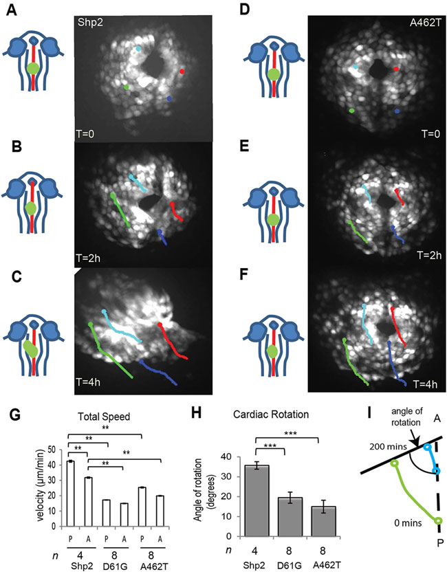
Impaired cardiomyocyte migration in embryos expressing Shp2-D61G and Shp2-A462T. Embryos from the tg(myl7:GFP) line were injected with mRNA encoding WT-Shp2, Shp2-D61G or Shp2-A462T lacking eGFP-peptide 2A sequences to allow imaging of GFP-positive cardiomyocytes from the 22-somite stage onwards. (A-F) Representative individual GFP-positive cells were color-coded according to their location within the cardiac field at the 22-somite stage and tracked over a 200 min period. Dorsal view with anterior towards the top. Schematic representations of the embryos on the left indicate the position and shape of the heart in green. (A-C) WT-Shp2; (D-F) Shp2-A462T. (G) Quantification of the total speed of posterior (P) and anterior (A) cardiac progenitor cells. WT-Shp2- expressing embryos displayed normal leftward heart displacement; embryos expressing Shp2-D61G and Shp2-A462T that did not display normal heart displacement were selected for further analysis. (H) Clockwise cardiomyocyte rotation was determined of the same embryos as in G. Rotation was quantified as depicted in I. (G,H) Averages are indicated and error bars indicate s.e.m.; n indicates number of embryos. Statistical significance was determined using Student’s t-test: **P<0.01 and ***P<0.001.