
|
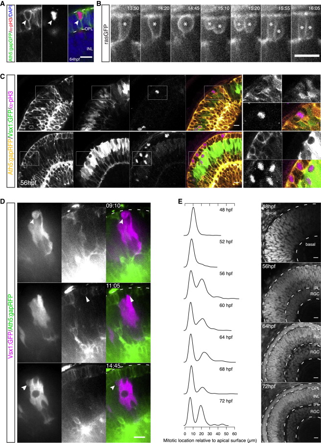
Fig. 3
Emergence of the Photoreceptor Cell Layer Leads to Repositioning of Bipolar Cell Precursor Divisions
(A) A pH3+/Ath5+ (red/green) cell within the PR layer (DAPI counterstaining in blue).
(B) A rasGFP+ cell (asterisk) undergoes division within the PR layer, giving rise to two daughters with PR-like morphology (from Movie S4). Time in hr:min. Imaging started at 50 hpf.
(C) An Ath5:gapRFP/Vsx1:GFP (yellow/green) embryo stained for pH3 (magenta) at 56 hpf. Mitoses of Vsx1+ cells occur apically in regions without a complete PR layer (top). In regions with an established PR layer, Vsx1+ divisions are shifted toward subapical locations (bottom). pH3+ cells at the apical side are Ath5+. Scale bar represents 25 μm.
(D) Time-lapse of Vsx1:GFP cells (magenta) transplanted into Ath5:gapRFP (green) background. Vsx1+ cells divide apically in regions without an established PR layer (top, notched arrow). Apical Ath5+ divisions can be observed in regions where PR precursors start to occupy the apical side (middle, filled arrow). Once a compact PR layer is established at the apical side, Vsx1+ divisions occur subapically (bottom, arrow). Time in hr:min. Imaging started at 44 hpf.
(E) Normalized kernel density estimates showing distributions of division locations in control embryos over time (left). Progression of retinal layer formation shown by DAPI staining (right). The emerging groove in the kernel density corresponds to the forming OPL.
If not stated differently, scale bars represent 10 μm. See also Figure S2.