
Fig. S7
- ID
- ZDB-IMAGE-140428-6
- Publication
- Hu et al., 2014 - Zebrafish eaf1 suppresses foxo3b expression to modulate transcriptional activity of gata1 and spi1 in primitive hematopoiesis
- All Figures
- Figures for Hu et al., 2014
|
Fig. S7
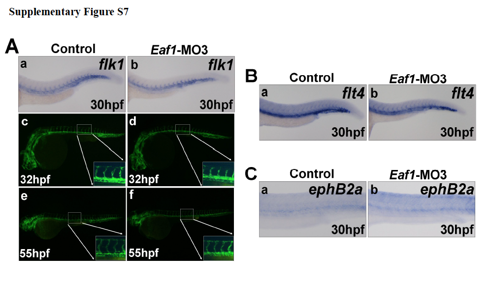
Knockdown of eaf1 does not disrupt blood vessel formation. (A) Expression of the vascular endothelial cell marker flk1 indicated a normal axial vasculature and initiation of inter-somatic vessels in eaf1 morphants (a-b). Wildtype transgenic embryos (flk1:EGFP) embryos at 32hpf (c). (d) Transgenic embryos (flk1::EGFP) injected with Eaf1-MO3 at 32hpf. Wildtype transgenic embryos (flk1::eGFP) at 55hpf (e). The overall vasculature was normally formed in eaf1-depleted embryos at 55hpf (f). (B) Expression of venous vessel marker flt4 in eaf1 morphants showed that the posterior cardinal vein was not affected by knockdown of eaf1. (C) Expression of arterial marker ephrinB2a in eaf1 morphants showed that the dorsal aorta was also not affected by the knockdown of eaf1. Eaf1-MO3, 4ng/embryo.
Reprinted from Developmental Biology, 388(1), Hu, B., Zhang, W., Feng, X., Ji, W., Xie, X., and Xiao, W., Zebrafish eaf1 suppresses foxo3b expression to modulate transcriptional activity of gata1 and spi1 in primitive hematopoiesis, 81-93, Copyright (2014) with permission from Elsevier. Full text @ Dev. Biol.