
Fig. 3
- ID
- ZDB-IMAGE-140416-46
- Genes
- Publication
- Uribe et al., 2013 - Ethanol affects the development of sensory hair cells in larval zebrafish (Danio rerio)
- All Figures
- Figures for Uribe et al., 2013
|
Fig. 3
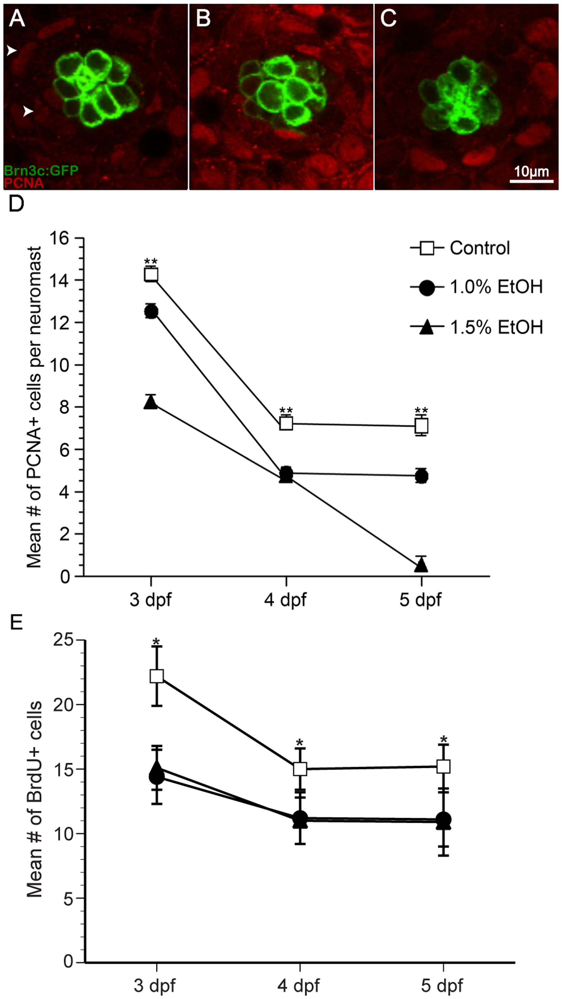
Ethanol exposure significantly reduced the number of proliferating cells compared to untreated controls in O2 neuromasts of Brn3c GFP-larvae.
(A) Under untreated control conditions, proliferating cell nuclear antigen (PCNA)-labeled cells (arrowhead) are primarily non-sensory supporting cells and mantle cells in the neuromast (area demarked by white dotted line). Fewer PCNA-labeled cells result from ethanol treatment at (B) 1% and (C) 1.5% by volume. (D) The mean number of PCNA-labeled cells decreases during maturation and according dose of ethanol. (E) In a separate experiment, bromodeoxyuridine was added to the embryo medium during the last 24 hours of treatment, fixed, processed for BrdU immunohistochemistry, and BrdU-labeled cells were counted. Significantly fewer BrdU-labeled cells were observed in ethanol-treated animals when compared to untreated controls. Results are the mean values ± SD. n = 10-21 neuromasts for each treatment group. **p<0.01.