
Fig. 5
- ID
- ZDB-IMAGE-140416-4
- Antibodies
- Publication
- Myhre et al., 2014 - The titin A-band rod domain is dispensable for initial thick filament assembly in zebrafish
- All Figures
- Figures for Myhre et al., 2014
|
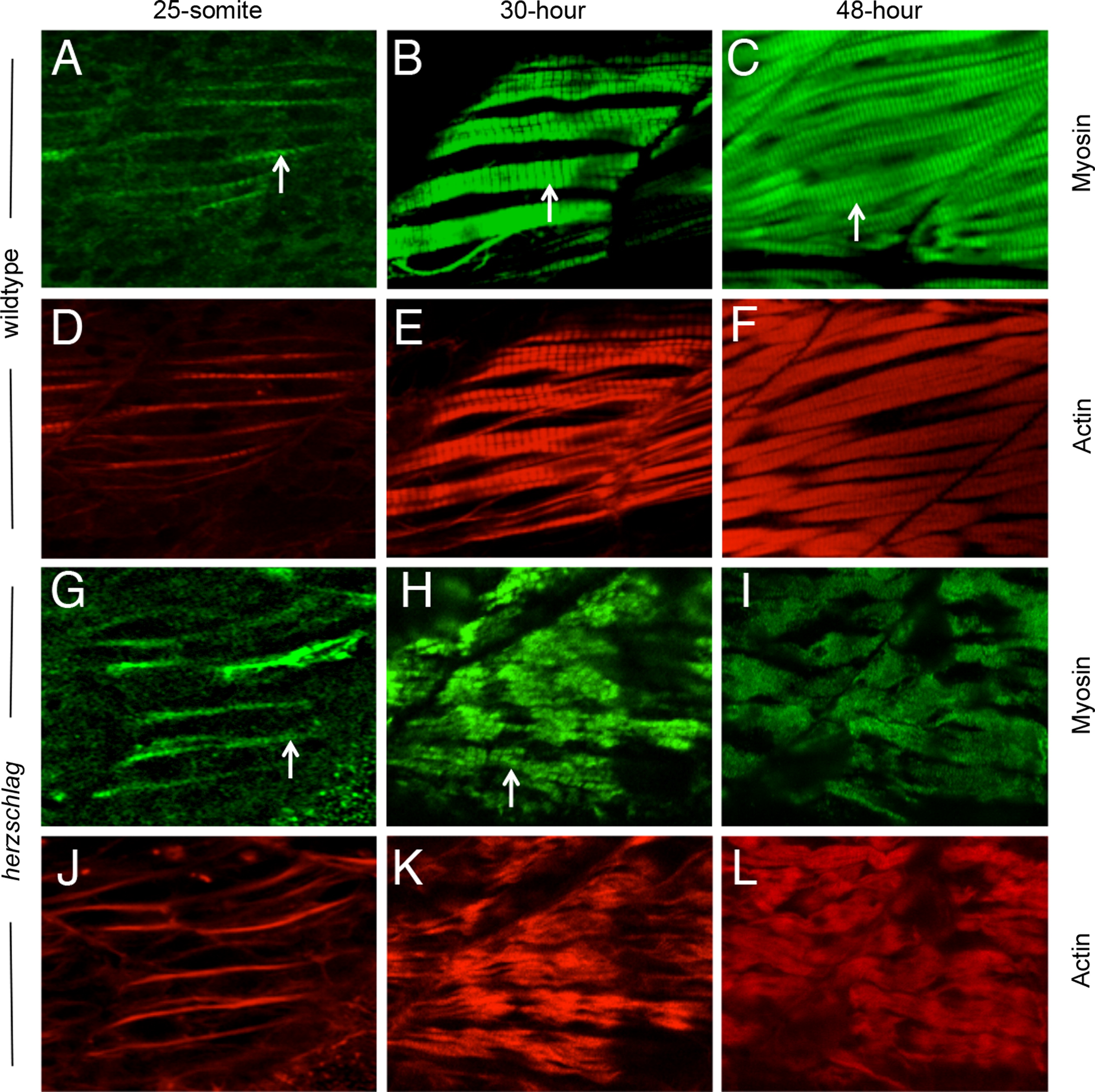
Fig. 5 Myosin thick filament organization still occurs in the absence of the titin rod domain. Herzschlag mutants and wild-type embryos were followed through a time-course of myogenesis, and stained at 24, 30 and 48 hpf with antibodies against slow-muscle myosin (green). In wild-type embryos (A–F), the striations of organized thick filaments can be seen at all time-points throughout myofibrillogenesis (arrows in A–C). Counter-staining with fluorescently labeled phalloidin to show actin organization (red) reveals a similar pattern of thin filament organization (D–F). In hel mutant embryos, the striated pattern of myosin thick filaments was retained at 24 h (G, arrow). By 30 hpf, myofibers were clearly disorganized (H), but retained a striated pattern of thick filament myosin (arrow). This pattern was only lost completely after 48 hpf (I). Thin filament striations, as show by phalloidin counter-staining, were never visible at any timepoint in hel mutant embryos (J–L). In all experiments, a minimum of 20 embryos were examined to ensure consistency of the phenotype.
Reprinted from Developmental Biology, 387(1), Myhre, J.L., Hills, J.A., Prill, K., Wohlgemuth, S.L., and Pilgrim, D.B., The titin A-band rod domain is dispensable for initial thick filament assembly in zebrafish, 93-108, Copyright (2014) with permission from Elsevier. Full text @ Dev. Biol.