
IMAGE
Fig. 2
- ID
- ZDB-IMAGE-140416-34
- Publication
- Viringipurampeer et al., 2014 - Rip3 knockdown rescues photoreceptor cell death in blind pde6c zebrafish
- All Figures
- Figures for Viringipurampeer et al., 2014
Image
|
Figure Caption
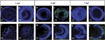
Fig. 2
Time course of TUNEL staining in whole-mount and retinal sections. Representative images of whole-mount eyes and corresponding sections at the denoted ages of 3, 4 or 7 days post fertilization (d.p.f.). (a, b, e, f, i, k) Whole-mount eyes. (c, d, g, h, k, l) Retinal sections. mt, pde6cw59/ mutant; wt, wild type. Scale bar is 20μm. TUNEL-positive cells, green labelling; 42,6-Diamidino-2-phenylindole counterstain for cell nuclei
Figure Data
Acknowledgments
This image is the copyrighted work of the attributed author or publisher, and
ZFIN has permission only to display this image to its users.
Additional permissions should be obtained from the applicable author or publisher of the image.
Full text @ Cell Death Differ.