
Fig. 1
- ID
- ZDB-IMAGE-140416-33
- Genes
- Publication
- Viringipurampeer et al., 2014 - Rip3 knockdown rescues photoreceptor cell death in blind pde6c zebrafish
- All Figures
- Figures for Viringipurampeer et al., 2014
|
Fig. 1
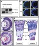
Genotyping and retinal histology for mutant pde6cw59 zebrafish. (a) Genotyping for mutation status using BsaX1 restriction digest of pde6c PCR amplicon. Wild-type (+/+) band is 157bp and the mutant (/) band is 122bp. (b) Heterozygous (+/) and mutant pde6c phenotype on transgenic background of GFP-expressing cone photoreceptors at 4 and 7 days post fertilization (d.p.f.) showing degeneration of cones. Scale bar=20μm. (c) Comparison of sections through heterozygous (+/) and mutant (–/–) retinas at 7>d.p.f. C, degenerating cones (black arrows); gcl, ganglion cell layer; inl, inner nuclear layer; onl, outer nuclear layer; r, single layer of rods (white arrowheads); black arrowheads, preserved peripheral retina. Scale bar=50μm. Dorsal retina is at the top of each panel. (d) Higher magnification of heterozygous (het) versus mutant (mt) retina at 4d.p.f. cn, cone nuclei; ipl, inner plexiform layer; rn, rod nuclei. Scale bar=μm. White arrows: condensed rod nuclei