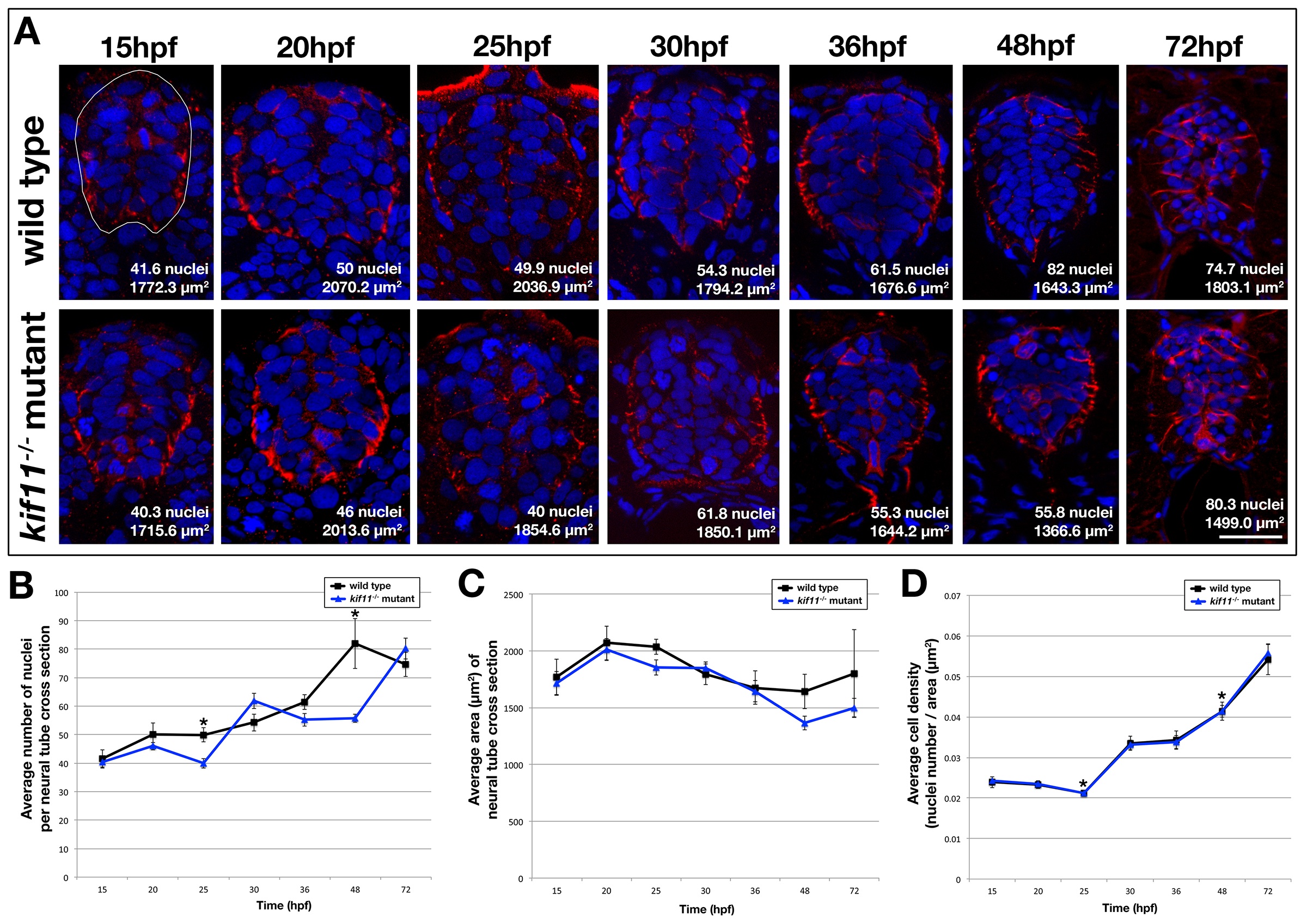
Fig. S3 Cell density in the kif11-/- neural tube is unchanged. (A) Cross sectional analysis of Gfap-positive astroglia (red) and Hoechst-positive nuclei (blue) in the developing spinal cord over time from 15hpf to 72hpf in wild type (top row) and kif11-/- mutants (bottom row). The cross sectional area of the neural tube was calculated for each section following manual outlining of the neural tube, which was based on Gfap labeling that delineates the outer edges of the neural tube (white line shown in 15hpf wild type image). (B-D) Quantification and data analysis of cell densities in the neural tube. Three sections corresponding to the axial locations of approximately somite 4-6, 10-12, and 16-18 were collected per embryo. 4 embryos were counted per time point. A resulting total of 12 sections were counted for 15-30hpf and 48hpf time points, and 11 sections were counted for 36hpf and 10 sections for 72hpf. Nuclei and area counts were averaged for each time point, and are shown on each example image (A). Only the 25hpf and 48hpf time points showed a statistically significant decrease in the number of nuclei in the kif11-/- mutant (blue line) neural tube as compared with wild type controls (black line) (B, asterisks). No statistically significant difference was seen in the cross sectional area of the neural tube between kif11 siblings (C). (D) Calculated cell density in the neural tube showed no visible difference between kif11 siblings (C, overlapping blue and black lines); however both 25hpf and 48hpf continued to be statistically significantly different (asterisks; 25hpf, p=0.004; 48hpf, p=0.004). Error bars delineate the standard error of the mean. Asterisks indicate a statistical significance (t-test, two tailed, assuming equal variances, p<0.05).
Reprinted from Developmental Biology, 387(1), Johnson, K., Moriarty, C., Tania, N., Ortman, A., Dipietrantonio, K., Edens, B., Eisenman, J., Ok, D., Krikorian, S., Barragan, J., Golé, C., and Barresi, M.J., Kif11 dependent cell cycle progression in radial glial cells is required for proper neurogenesis in the zebrafish neural tube, 73-92, Copyright (2014) with permission from Elsevier. Full text @ Dev. Biol.

