
Image
|
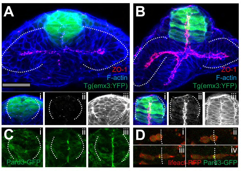
Figure Caption
Fig. S3
Time-lapse movie sequence of eye field core cells expressing the fusion protein Pard3-GFP (green). The embryo is counter-labeled with a membrane-RFP (red). Insets below show the green channel only (Pard3-GFP). A subset of the labelled core cells coordinate their subcellular polarised localisation of Pard3-GFP and organise as a rosette-like structure (follow the yellow arrow, n=5 in 4 embryos). Some others did not do so (red arrow).
Acknowledgments
This image is the copyrighted work of the attributed author or publisher, and
ZFIN has permission only to display this image to its users.
Additional permissions should be obtained from the applicable author or publisher of the image.
Reprinted from Developmental Cell, 27(3), Ivanovitch, K., Cavodeassi, F., and Wilson, S.W., Precocious acquisition of neuroepithelial character in the eye field underlies the onset of eye morphogenesis, 293-305, Copyright (2013) with permission from Elsevier. Full text @ Dev. Cell