
|
Fig. S1
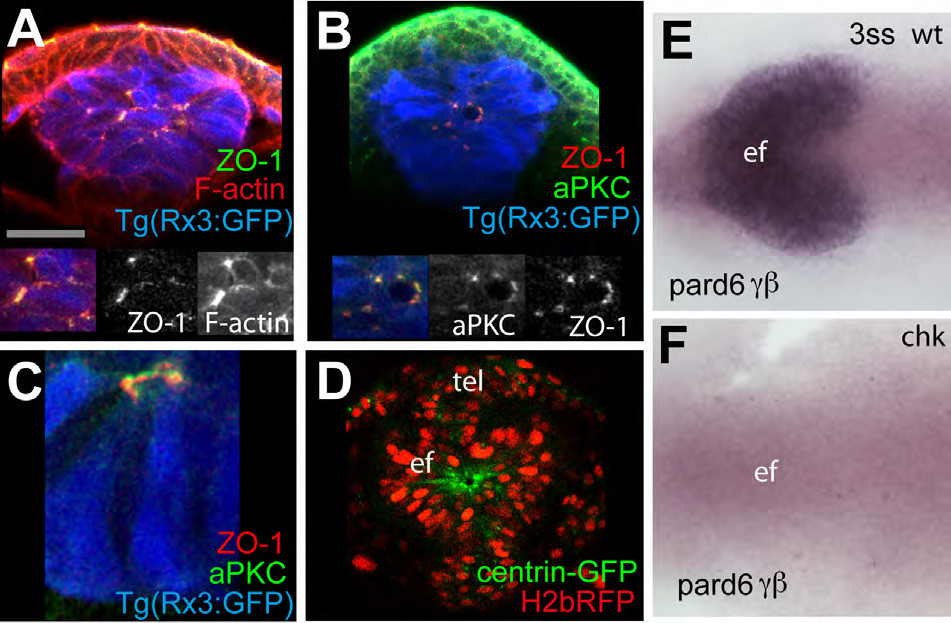
(A-C) 4ss Tg(rx3:GFP) embryos immunostained as detailed in the panels [ZO-1 (green in A, red in B and C), F-actin (red, A) and aPKC (green in B)]. Inset boxes showing F-actin and aPKC, and ZO- 1 and aPKC colocalise in the eye field at 4ss. aPKC can sometimes localise apically to ZO-1 (C). (D) View of the eyefield of a wildtype embryo injected with mRNA for H2b-RFP to label the nuclei and centrin-GFP to label centrosomes, showing accumulation of this marker at the centre of the eye field. White dotted lines in (D) outline the eye field. tel: telencephalon; ef: eye field. (E-F) in situ hybridization to detect pard6γβ in the eye field of wildtype (E) and rx3chk mutant embryos (F).
Reprinted from Developmental Cell, 27(3), Ivanovitch, K., Cavodeassi, F., and Wilson, S.W., Precocious acquisition of neuroepithelial character in the eye field underlies the onset of eye morphogenesis, 293-305, Copyright (2013) with permission from Elsevier. Full text @ Dev. Cell